Salivary gland mucocele
Jump to navigation
Jump to search
Salivary gland mucocele, also salivary mucocele and mucocele, is a benign lesion of the head and neck.
General
- Benign.
- Infected mucocele = mucopyocele.
Gross
Features:
- Floor of mouth lesion.
Images
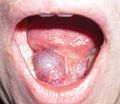
Ranula. (WC/Ph0t0happy)

Microscopic
Features:[1]
- Granulation tissue-like and pseudocyst-like.
- Granulation tissue-like:
- Fibroblasts.
- Small caliber blood vessels.
- Histocytes.
- Neutrophils.
- Pseudocyst:
- No epithelial lining.
- Poorly circumscribed.
- Granulation tissue-like:
- Pale pink extracellular material (mucous) - key feature.
- +/-Granulomas.[2]
DDx:
- Granulation tissue.
- Signet ring cell carcinoma - muciphages may mimic signet ring cells.
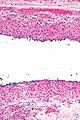
Images
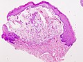
Mucocele - low mag. (WC/KGH)
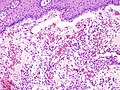
Mucocele - high mag. (WC/KGH)
Mucocele - low mag. (WC/Nephron)
Mucocele - high mag. (WC/Nephron)
.JPG)
.JPG)


www:
Sign out
LESION, LEFT LOWER LIP, EXCISION:
- BENIGN MUCOCELE.
Micro
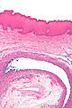
The sections show a stratified squamous epithelium with a thin layer of parakeratosis, minor salivary glands, and a well-circumscribed cystic lesion.
The cystic lesion has a mildly fibrotic appearing wall, is lined by histiocytes intermixed with rare lymphocytes, and contains mucous and macrophages. No significant nuclear atypia is identified. Mitotic activity is not readily apparent.
See also
References
- ↑ URL: http://emedicine.medscape.com/article/1076717-workup. Accessed on: 6 March 2012.
- ↑ Seifert, G.; Donath, K.; von Gumberz, C. (Jun 1981). "[Mucoceles of the minor salivary glands. Extravasation mucoceles (mucus granulomas) and retention mucoceles (mucus retention cysts) (author's transl)].". HNO 29 (6): 179-91. PMID 7251405.