Rheumatoid arthritis
Jump to navigation
Jump to search
Rheumatoid arthritis, commonly abbreviated RA, is an autoimmune disorder.
Skin
Rheumatoid neutrophilic dermatitis
General
- Case report rare manifestation of rheumatoid arthritis.[1]
Microscopic
Features:
- Nodular and diffuse pattern.
- Neutrophils - perivascular (without vessel wall injury).[1]
DDx:
Joints
Main article: Joints
Rheumatic joint disease
- AKA joint with rheumatic disease.
General
Clinical:
- Tumour - swelling.
- Rubor - redness.
- Calor - heat.
- Dolor - pain.
Gross
Features:
- Pannus[2] - fibrovascular tissue or granulation tissue.
- Irregular surface - synovial hyperplasia.
- Subchondral cysts - involve the entire femoral head (late stage of disease).[3]
Note:
- Osteoarthritis subchondral cysts are associated with cartilage loss.[3]
Image:
Microscopic
Features:[5]
- Chronic inflammation, esp. lymphocytes.
- +/-Lymphoid follicles.
- Synovial hyperplasia - with papillary or polypoid architecture.
- Synoviocytes may show binucleation and mild atypia.
- +/-Fibrin.
- +/-Bone.
- +/-Cartilage.
Note:
- Changes are non-specific - DDx includes other rheumatic diseases (systemic lupus erythematosus, psoriatic arthritis).
DDx:
- Infected joint.
Images:
- RA (med.utah.edu).[6]
- Rheumatoid arthritis (nature.com).
- Synovial inflammation (medpath.info).[4]
- Hyperplastic synovitis (medpath.info).[4]
- RA cartilage loss (bmj.com).
Sign out
Femoral head
FEMORAL HEAD, LEFT, HIP ARTHROPLASTY:
- CHRONIC SYNOVITIS WITH SYNOVIAL HYPERPLASIA AND LOSS OF CARTILAGE.
- BONE WITHOUT APPARENT PATHOLOGY.
- SEE COMMENT.

COMMENT:
The findings are compatible with rheumatoid arthritis.
Hand
SYNOVIUM, LEFT HAND, EXCISION:
- SYNOVIAL HYPERPLASIA.
- CHRONIC AND FOCAL ACUTE SYNOVITIS.
- FIBRINOUS EXUDATE.
- GRANULATION TISSUE AND HEMOSIDERIN-LADEN MACROPHAGES.
- SEE COMMENT.

COMMENT:
The findings are compatible with rheumatoid arthritis.
Knee
KNEE - BONE AND SOFT TISSUE, RIGHT, KNEE ARTHROPLASTY:
- CHRONIC SYNOVITIS WITH SYNOVIAL HYPERPLASIA AND LOSS OF CARTILAGE.
- BONE WITHOUT APPARENT PATHOLOGY.

COMMENT:
The findings are compatible with rheumatoid arthritis.
KNEE - BONE AND SOFT TISSUE, LEFT, KNEE ARTHROPLASTY:
- DEGENERATIVE JOINT DISEASE WITH CHRONIC SYNOVITIS AND SYNOVIAL HYPERPLASIA.
- BONE WITHOUT APPARENT PATHOLOGY.

COMMENT:
The findings are compatible with rheumatoid arthritis.
Rheumatoid nodule
| Rheumatoid nodule | |
|---|---|
| External resources | |
| EHVSC | 10182 |
- Abbreviated RN.
General
- Seen in rheumatoid arthritis - usually only in seropositive cases, i.e. rheumatoid factor (RF) positive.[7]
- Exceptions are reported.[8]
Gross
- Usually on the extensor aspect of the extremities, e.g. dorsal aspect of elbow.[9]
- Typically close to a joint.[citation needed]
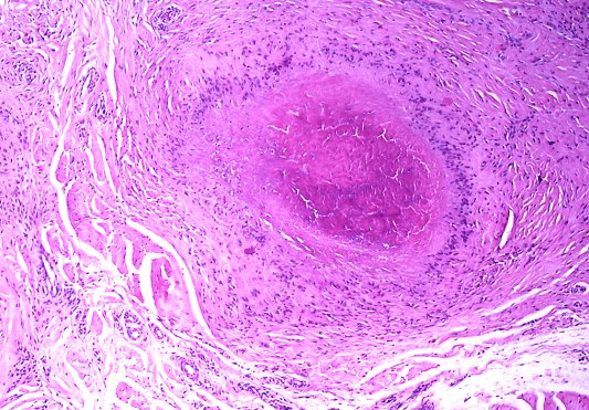
Microscopic
- Necrotic collagen bundles with fibrin surrounded by:
- Palisading granuloma.
- +/-Eosinophils.
Notes:
- Histomorphologically very similar to Granuloma annulare.
DDx:
- Granuloma annulare - has mucin in the core of the granuloma.[10]
- Necrobiosis lipoidica.
Images
www:
- Rheumatoid nodule (granuloma.homestead.com).[11]
- Rheumatoid nodule (granuloma.homestead.com).[11]
- Rheumatoid nodule (utah.edu).[12]
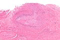
Rheumatoid nodule - low mag. (WC)
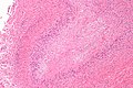
Rheumatoid nodule - intermed. mag. (WC)
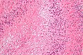
Rheumatoid nodule - high mag. (WC)


Stains
- Alcian blue (pH 2.5) -ve.
- Positive staining of mucin in granuloma annulare.[13]
Sign out
Lesion (subcutaneous), right index finger, excision:
- Palisading granulomas with cores of necrobiotic collagen, consistent with rheumatoid nodule.
Micro
The sections show foci with strands of necrobiotic collagen surrounded by palisading granulomas. Rare Neutrophils are seen in the core of the granulomas. Multinucleated cells are not apparent.
Benign fibroadipose tissue and dense connective tissue are also present.
Pleural disease
- See Rheumatoid pleuritis.
Lung disease
- See Medical lung disease.
RA may involve the lung.
Miscellaneous
Amyloidosis
- See Amyloidosis.
Amyloidosis may be due to RA.
Felty syndrome
RA may occur in Felty syndrome -- the triad:[14]
- Rheumatoid arthritis.
- Splenomegaly.
- Neutropenia.
Felty syndrome is associated with large granular lymphocytic leukemia.[14][15]
See also
References
- ↑ 1.0 1.1 Mashek, HA.; Pham, CT.; Helm, TN.; Klaus, M. (Jun 1997). "Rheumatoid neutrophilic dermatitis.". Arch Dermatol 133 (6): 757-60. PMID 9197831.
- ↑ Lester, Susan Carole (2005). Manual of Surgical Pathology (2nd ed.). Saunders. pp. 223. ISBN 978-0443066450.
- ↑ 3.0 3.1 Resnick, D.; Niwayama, G.; Coutts, RD. (May 1977). "Subchondral cysts (geodes) in arthritic disorders: pathologic and radiographic appearance of the hip joint.". AJR Am J Roentgenol 128 (5): 799-806. PMID 404905.
- ↑ 4.0 4.1 4.2 URL: http://www.medpath.info/MainContent/Skeletal/Joint_02.html. Accessed on: 10 November 2012.
- ↑ Humphrey, Peter A; Dehner, Louis P; Pfeifer, John D (2008). The Washington Manual of Surgical Pathology (1st ed.). Lippincott Williams & Wilkins. pp. 660. ISBN 978-0781765275.
- ↑ URL: http://library.med.utah.edu/WebPath/EXAM/IMGQUIZ/msfrm.html. Accessed on: 5 December 2010.
- ↑ 7.0 7.1 Tadrous, Paul.J. Diagnostic Criteria Handbook in Histopathology: A Surgical Pathology Vade Mecum (1st ed.). Wiley. pp. 299. ISBN 978-0470519035.
- ↑ Kaye, BR.; Kaye, RL.; Bobrove, A. (Feb 1984). "Rheumatoid nodules. Review of the spectrum of associated conditions and proposal of a new classification, with a report of four seronegative cases.". Am J Med 76 (2): 279-92. PMID 6364806.
- ↑ Busam, Klaus J. (2009). Dermatopathology: A Volume in the Foundations in Diagnostic Pathology Series (1st ed.). Saunders. pp. 53. ISBN 978-0443066542.
- ↑ 10.0 10.1 Busam, Klaus J. (2009). Dermatopathology: A Volume in the Foundations in Diagnostic Pathology Series (1st ed.). Saunders. pp. 52. ISBN 978-0443066542.
- ↑ 11.0 11.1 URL: http://granuloma.homestead.com/palisading.html. Accessed on: 1 November 2010.
- ↑ URL: http://www.pathguy.com/lectures/joints.htm. Accessed on: 1 November 2010.
- ↑ Yun, JH.; Lee, JY.; Kim, MK.; Seo, YJ.; Kim, MH.; Cho, KH.; Kim, MB.; Lee, WS. et al. (May 2009). "Clinical and pathological features of generalized granuloma annulare with their correlation: a retrospective multicenter study in Korea.". Ann Dermatol 21 (2): 113-9. doi:10.5021/ad.2009.21.2.113. PMC 2861218. PMID 20523767. https://www.ncbi.nlm.nih.gov/pmc/articles/PMC2861218/.
- ↑ 14.0 14.1 Mitchell, Richard; Kumar, Vinay; Fausto, Nelson; Abbas, Abul K.; Aster, Jon (2011). Pocket Companion to Robbins & Cotran Pathologic Basis of Disease (8th ed.). Elsevier Saunders. pp. 328. ISBN 978-1416054542.
- ↑ Liu, X.; Loughran, TP. (Jul 2011). "The spectrum of large granular lymphocyte leukemia and Felty's syndrome.". Curr Opin Hematol 18 (4): 254-9. doi:10.1097/MOH.0b013e32834760fb. PMID 21546829.