Retinoblastoma
Jump to navigation
Jump to search
The printable version is no longer supported and may have rendering errors. Please update your browser bookmarks and please use the default browser print function instead.
| Retinoblastoma | |
|---|---|
| Diagnosis in short | |
 Retinoblastoma. (WC) | |
|
| |
| LM | small round cell tumour, Flexner-Wintersteiner rosettes, +/-Homer-Wright rosettes, mitoses (common), +/-necrosis, +/-calcifications |
| LM DDx | retinocytoma, other small round cell tumours |
| Gross | white, solid, friable |
| Site | eye |
|
| |
| Prevalence | rare |
Retinoblastoma is a malignant tumour the of the eye.
General
Gross
- White, solid.
- Patterns:
- Endophytic - grow into the vitreous cavity.
- Exophytic - grow toward choroid.
- Mixed - components of endophytic and exophytic.
Note:
- Tumour is extremely friable.
Image
Microscopic
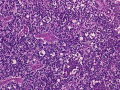
Features:
- Small round cell tumour:
- Scant cytoplasm.
- Flexner-Wintersteiner rosette - key feature.
- Rosette with empty centre (donut hole).[2]
- +/-Homer-Wright rosette.[3]
- Circular rosette with neuropil at the centre.[2]
- Mitoses - common.
- +/-Necrosis.
- +/-Calcification.
DDx:
- Retinocytoma (retinoma) - benign counterpart of retinoblastoma.
Notes:
- DDx of Flexner-Wintersteiner rosette includes:
- Pineoblastoma.
- Medulloepithelioma.
Images
Retinoblastoma. (WC)