Difference between revisions of "Renomedullary interstitial cell tumour"
Jump to navigation
Jump to search
(+infobox) |
(→Images) |
||
| Line 54: | Line 54: | ||
Image:Renal_medullary_fibroma_-_low_mag.jpg | Renal medullary fibroma - low mag. (WC/Nephron) | Image:Renal_medullary_fibroma_-_low_mag.jpg | Renal medullary fibroma - low mag. (WC/Nephron) | ||
Image:Renal_medullary_fibroma_-_intermed_mag.jpg | Renal medullary fibroma - intermed. mag. (WC/Nephron) | Image:Renal_medullary_fibroma_-_intermed_mag.jpg | Renal medullary fibroma - intermed. mag. (WC/Nephron) | ||
Image:Renal_medullary_fibroma_-_high_mag.jpg | Renal medullary fibroma - high mag. (WC/Nephron) | |||
Image:Renal_medullary_fibroma_-_very_high_mag.jpg | Renal medullary fibroma - very high mag. (WC/Nephron) | Image:Renal_medullary_fibroma_-_very_high_mag.jpg | Renal medullary fibroma - very high mag. (WC/Nephron) | ||
</gallery> | </gallery> | ||
| Line 59: | Line 60: | ||
*[http://webpathology.com/image.asp?case=71&n=15 Renomedullary interstitial cell tumour - low mag. (webpathology.com)]. | *[http://webpathology.com/image.asp?case=71&n=15 Renomedullary interstitial cell tumour - low mag. (webpathology.com)]. | ||
*[http://webpathology.com/image.asp?n=16&Case=71 Renomedullary interstitial cell tumour - high mag. (webpathology.com)]. | *[http://webpathology.com/image.asp?n=16&Case=71 Renomedullary interstitial cell tumour - high mag. (webpathology.com)]. | ||
==See also== | ==See also== | ||
*[[Kidney tumours]]. | *[[Kidney tumours]]. | ||
Revision as of 03:29, 22 December 2013
| Renomedullary interstitial cell tumour | |
|---|---|
| Diagnosis in short | |
 Renal medullary fibroma. H&E stain. | |
|
| |
| Synonyms | renal medullary fibroma |
|
| |
| LM | small polygonal/stellate cells in abundant loose/myxoid stroma, +/-entrapped renal tubules |
| Gross | small (usu. <3 mm), white, well-circumscribed nodule - medulla of kidney |
| Site | kidney - see kidney tumours |
|
| |
| Prevalence | common |
| Prognosis | benign |
Renomedullary interstitial cell tumour, also known as medullary fibroma,[1] is a relatively common benign tumour of the kidney.
General
- Benign.
- Common autopsy finding[2] - one review says 26-41% of individuals at autopsy.[3]
- The commonality is somewhat in dispute.[4]
Gross
- Small, white well-circumscribed nodule in medulla.
- Typically less than 3 mm.[3]
Image:
Microscopic
- Small polygonal/stellate cells.
- Abundant loose/myxoid stroma.
- +/-Entrapped renal tubules.[6]
Images
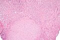
Renal medullary fibroma - low mag. (WC/Nephron)
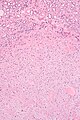
Renal medullary fibroma - intermed. mag. (WC/Nephron)
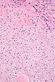
Renal medullary fibroma - high mag. (WC/Nephron)
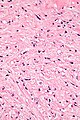
Renal medullary fibroma - very high mag. (WC/Nephron)


www:
- Renomedullary interstitial cell tumour - low mag. (webpathology.com).
- Renomedullary interstitial cell tumour - high mag. (webpathology.com).
See also
References
- ↑ Bircan, S.; Orhan, D.; Tulunay, O.; Safak, M. (2000). "Renomedullary interstitial cell tumor.". Urol Int 65 (3): 163-6. PMID 11054036.
- ↑ 2.0 2.1 Humphrey, Peter A; Dehner, Louis P; Pfeifer, John D (2008). The Washington Manual of Surgical Pathology (1st ed.). Lippincott Williams & Wilkins. pp. 295. ISBN 978-0781765275.
- ↑ 3.0 3.1 Tsurukawa, H.; Iuchi, H.; Osanai, H.; Yamaguchi, S.; Hashimoto, H.; Kaneko, S.; Yachiku, S. (Jan 2000). "[Renomedullary interstitial cell tumor: a case report].". Nihon Hinyokika Gakkai Zasshi 91 (1): 37-40. PMID 10689882.
- ↑ Kozłowska, J.; Okoń, K. (2008). "Renal tumors in postmortem material.". Pol J Pathol 59 (1): 21-5. PMID 18655367.
- ↑ URL: http://webpathology.com/image.asp?n=16&Case=71. Accessed on: 17 October 2011.
- ↑ Kuroda, N.; Toi, M.; Miyazaki, E.; Hayashi, Y.; Nakayama, H.; Hiroi, M.; Enzan, H.. "Participation of alpha-smooth muscle actin-positive cells in renomedullary interstitial cell tumors.". Oncol Rep 9 (4): 745-50. PMID 12066202.