Pulmonary infarct
Jump to navigation
Jump to search
The printable version is no longer supported and may have rendering errors. Please update your browser bookmarks and please use the default browser print function instead.
| Pulmonary infarct | |
|---|---|
| Diagnosis in short | |
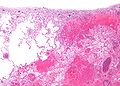
 Pulmonary infarct. H&E stain. | |
|
| |
| Synonyms | lung infarct |
|
| |
| LM | necrosis of alveolar walls - loss of nuclei, alveolar hemorrhage, +/-evidence of underlying cause |
| LM DDx | see Associated Dx |
| Gross | lung periphery, classically described as wedge-shaped |
| Site | lung |
|
| |
| Associated Dx | underlying causes: sickle cell disease, pulmonary embolism, vasculitides, malignancy (e.g. lymphoma), drug toxicity, others |
| Prevalence | uncommon |
| Radiology | reverse halo sign |
| Prognosis | dependent on underlying cause |
| Treatment | dependent on underlying cause |
Pulmonary infarct is the death of lung tissue due to oxygen deprivation.
It is also known as a lung infarct, lung infarction, and pulmonary infarction.
General
- Uncommon because of the dual blood supply (systemic via the bronchial arteries, pulmonary via the pulmonary arteries).
Common causes:[1]
Less common causes:
- Lymphoma, esp. acute promyelocytic leukemia.
- Drugs, e.g. chemotherapy.
- Vasculitis.
- Others.
Gross
- Lung periphery, classically described as wedge-shaped.
Note:
- In a histologic section, the classic wedge-shaped infarct is triangular:
- Base of triangle on the pleural aspect.
- Point furthest from the pleura close to the compromised artery that lead to infarction.
Radiology:
- Reverse halo sign.[2]
Images:
Microscopic
Features:
- Necrosis of alveolar walls - loss of nuclei.
- Alveolar hemorrhage.
Image
Pulmonary infarct - low mag. (WC)
See also
References
- ↑ URL: http://emedicine.medscape.com/article/908045-overview. Accessed on: 12 April 2012.
- ↑ 2.0 2.1 Casullo, J.; Semionov, A. (Feb 2013). "Reversed halo sign in acute pulmonary embolism and infarction.". Acta Radiol. doi:10.1177/0284185113475797. PMID 23395814.