Pulmonary carcinoid tumourlet
Jump to navigation
Jump to search
The printable version is no longer supported and may have rendering errors. Please update your browser bookmarks and please use the default browser print function instead.
| Pulmonary carcinoid tumourlet | |
|---|---|
| Diagnosis in short | |
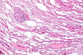
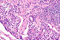
 Lung carcinoid tumourlet. H&E stain. | |
|
| |
| Synonyms | carcinoid tumourlet |
|
| |
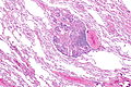
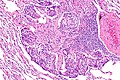
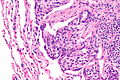
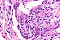
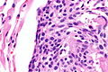
| LM | cells with salt and pepper chromatin, usually nested architecture, no necrosis, minimal mitotic activity (see below), must be through the bronchial basement membrane |
| LM DDx | pulmonary neuroendocrine cell hyperplasia, typical carcinoid lung tumour, atypical lung carcinoid tumour, pulmonary meningothelial-like nodule |
| IHC | Ki-67 ~2% (0-7%) |
| Gross | <5 mm by definition |
| Site | lung - see lung tumours |
|
| |
| Syndromes | Diffuse idiopathic pulmonary neuroendocrine cell hyperplasia |
|
| |
| Clinical history | often an incidental finding |
| Prevalence | not common |
| Prognosis | benign |
Pulmonary carcinoid tumourlet, also carcinoid tumourlet, is a small benign proliferation of Kulchitsky cells.
The entity is separated from the typical lung carcinoid tumour by size. Carcinoid tumourlets are < 5 mm, typical lung carcinoid tumours are >=5 mm.
General
- Neuroendocrine cell proliferation.[1]
- Essentially a small typical carcinoid.
- Arise from Kulchitsky cells of the bronchial epithelium.[2]
- May be seen in the context of diffuse idiopathic pulmonary neuroendocrine cell hyperplasia.
Microscopic
Features:
- Neuroendocrine cells - usually in nests (classic pattern).
- Salt and pepper chromatin - key feature.
- Nuclei round or ellipsoid.
- Size criterion: <5 mm.[3][4]
- Must be through the bronchial basement membrane.[5]
DDx:
- Pulmonary neuroendocrine cell hyperplasia - proliferation confined by bronchial basement membrane.[5]
- Typical carcinoid lung tumour - must be >= 5 mm.
- Pulmonary meningothelial-like nodule - whorled appearance, not associated with an airway.
Images
CT - very low mag. (WC)
CT - low mag. (WC)
CT - intermed. mag. (WC)
CT - high mag. (WC)
CT - high mag. (WC)
CT - very high mag. (WC)
CT - very high mag. (WC)


www:
Sign out
A. Lymph Node, Station 4R, Lymphadenectomy:
- Lymph node, NEGATIVE for malignancy.

B. Lymph Node, Station 11R, Lymphadenectomy:
- Lymph node, NEGATIVE for malignancy.

C. Lung, Right Middle Lobe, Lobectomy:
- Typical carcinoid tumour (13 mm maximal dimension).
- Carcinoid tumourlet (3 mm maximal dimension).
- Margins clear of tumour.
- Please see tumour summary.
See also
References
- ↑ Bennett, GL.; Chew, FS. (Mar 1994). "Pulmonary carcinoid tumorlets.". AJR Am J Roentgenol 162 (3): 568. PMID 8109497.
- ↑ Ramón Capilla, M.; Arnau Obrer, A.; Navarro Ibáñez, R.; Galbis Caravajal, J.; Traves Zapata, V.; Cantó Armengod, A. (Nov 1996). "[Pulmonary tumorlet. Report of 5 cases].". Arch Bronconeumol 32 (9): 489-91. PMID 9064089.
- ↑ URL: http://pathhsw5m54.ucsf.edu/case7/image75.html. Accessed on: 23 January 2012.
- ↑ He, P.; Gu, X.; Wu, Q.; Lin, Y.; Gu, Y.; He, J. (Dec 2012). "Pulmonary carcinoid tumorlet without underlying lung disease: analysis of its relationship to fibrosis.". J Thorac Dis 4 (6): 655-8. doi:10.3978/j.issn.2072-1439.2012.06.11. PMID 23205296.
- ↑ 5.0 5.1 Koo, CW.; Baliff, JP.; Torigian, DA.; Litzky, LA.; Gefter, WB.; Akers, SR. (Sep 2010). "Spectrum of pulmonary neuroendocrine cell proliferation: diffuse idiopathic pulmonary neuroendocrine cell hyperplasia, tumorlet, and carcinoids.". AJR Am J Roentgenol 195 (3): 661-8. doi:10.2214/AJR.09.3811. PMID 20729444.