Difference between revisions of "Primary biliary cholangitis"
Jump to navigation
Jump to search
m (replace) |
(→Images) |
||
| Line 85: | Line 85: | ||
www: | www: | ||
*[http://www.gidesigns.net/images/MC-copper-flower-garland-L.jpg Garland - wreath of flowers (gidesigns.net)]. | *[http://www.gidesigns.net/images/MC-copper-flower-garland-L.jpg Garland - wreath of flowers (gidesigns.net)]. | ||
{| | |||
[[File:1 PBC - 1 - 40X 680x512px.tif|Acini & lobules distorted by inflammation]] | |||
[[File:2 PBC - 1 - 200X 680x512px.tif|Loose granuloma in triad without duct]] | |||
[[File:3 PBC - 1 - 200X 680x512px.tif|Well-formed granuloma]] | |||
[[File:4 PBC - 1 -200X 680x512px.tif|Piecemeal necrosis, PAS without diastase]] | |||
[[File:5 PBC - 1 -400X 680x512px.tif|Damaged bile duct, PAS with diastase]] | |||
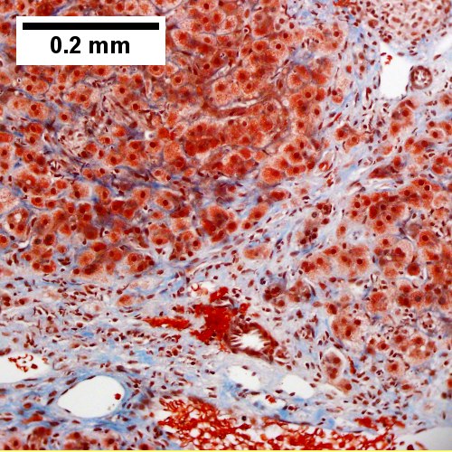
[[File:6 PBC - 1 -200X 680x512px.tif|Bridging fibrosis, trichrome]] | |||
|} | |||
Primary biliary cirrhosis with bridging fibrosis. Acini & lobules distorted by inflammation (TL,40X) Loose granuloma in triad without duct (TR,200X) Well-formed granuloma (ML,200X) Piecemeal necrosis, PAS without diastase (MR,200X) Damaged bile duct, PAS with diastase (BL,400X) Bridging fibrosis, trichrome (BR,200X) | |||
===Staging PBC=== | ===Staging PBC=== | ||
Revision as of 21:59, 7 July 2016
| Primary biliary cholangitis | |
|---|---|
| Diagnosis in short | |
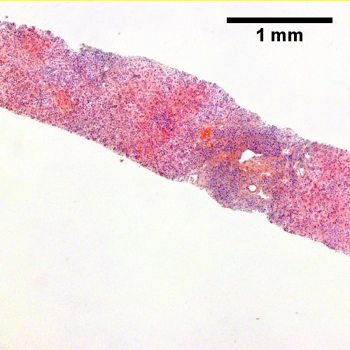
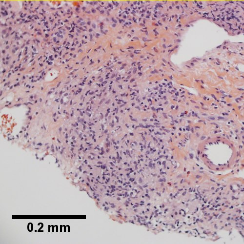
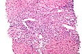
 Primary biliary cirrhosis. H&E stain. | |
|
| |
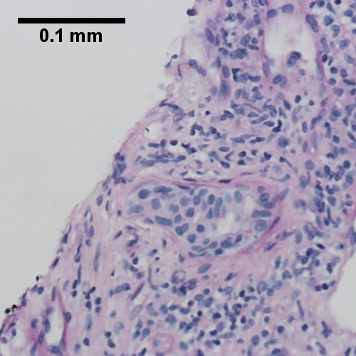
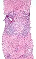
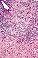
| LM | "florid duct lesion": intraepithelial lymphocytes - in bile duct, bile duct epithelial cells with eosinophilic cytoplasm; plasma cells; +/-granulomas (close to bile duct); +/-"garland" cirrhosis -- has irregular border |
| LM DDx | sarcoidosis, primary sclerosing cholangitis, viral hepatitis, autoimmune hepatitis, drug-induced liver disease, Hodgkin's lymphoma |
| Site | liver - see medical liver diseases |
|
| |
| Associated Dx | other autoimmune conditions, e.g. celiac disease, Sjögren syndrome |
| Clinical history | woman, middle age |
| Symptoms | pruritis |
| Blood work | AMA +ve |
Primary biliary cirrhosis, abbreviated PBC, is a rare medical liver disease.
General
Epidemiology:
- Female>male (~9:1).[1]
- Usually middle age.
- Associated with other autoimmune conditions (Sjögren syndrome, progressive systemic sclerosis, celiac disease).
Etiology:
- Autoimmune.
Serology:
- AMA +ve.[2]
Classic presentation:
- Pruritis.
Pathophysiology:
- Septal bile duct attacked.
Treatment:
- Ursodeoxycholic acid.
- May be indication for transplant.
Microscopic
Features:
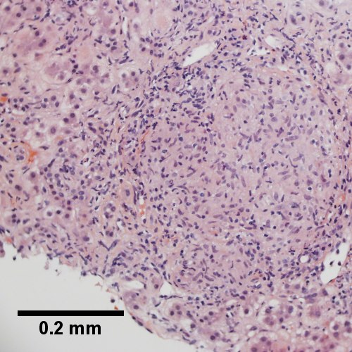
- "Florid duct lesion":[3]
- Intraepithelial lymphocytes - in bile duct - key feature.
- Bile duct epithelial cells with eosinophilic cytoplasm.[4]
- Plasma cells.
- Granulomas - close to bile duct.
- Seen in classic presentation -- often not present or poorly formed.
- Focal damage (may be missed on biopsy -- due to sampling).
- "Garland" cirrhosis -- has irregular border (unlike in EtOH).
- Garland originally "wreath of flowers" (in French).[5]
Notes:
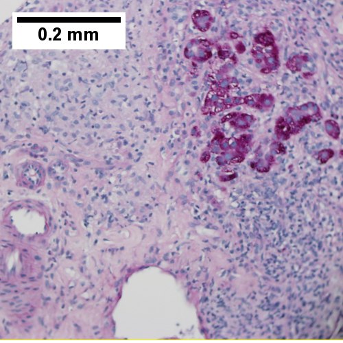
- PAS stain useful for examining basement membrane... which is lost in PBC.
- Lobular inflammation should be minimal.
- May cause cholestatic picture.[6]
DDx:[7]
- Sarcoidosis (if granulomas present).
- Primary sclerosing cholangitis.
- Viral hepatitis.
- Autoimmune hepatitis.
- Drug-induced liver disease.
- Hodgkin's lymphoma.[8]
Images
PBC - low mag. (WC)
PBC - intermed. mag. (WC)
PBC - intermed. mag. (WC)


www:
Primary biliary cirrhosis with bridging fibrosis. Acini & lobules distorted by inflammation (TL,40X) Loose granuloma in triad without duct (TR,200X) Well-formed granuloma (ML,200X) Piecemeal necrosis, PAS without diastase (MR,200X) Damaged bile duct, PAS with diastase (BL,400X) Bridging fibrosis, trichrome (BR,200X)
Staging PBC
PBC is staged according to Ludwig:[9]
- Stage 1: Portal - inflammation or bile duct abnormalities.
- Stage 2: Periportal - periportal fibrosis (enlargement of portal tracts) +/- inflammation.
- Stage 3: Septal - septal fibrosis +/-inflammation in septa.
- Stage 4: Cirrhosis - nodules of hepatocytes +/- inflammation.
Notes:
- There can be significant variation in staging on biopsy - due to variability of fibrosis in a PBC liver.[10]
- "Worst area" in biopsy specimen is used to determine stage.
See also
References
- ↑ Tadrous, Paul.J. Diagnostic Criteria Handbook in Histopathology: A Surgical Pathology Vade Mecum (1st ed.). Wiley. pp. 162. ISBN 978-0470519035.
- ↑ Nguyen, DL.; Juran, BD.; Lazaridis, KN. (Oct 2010). "Primary biliary cirrhosis.". Best Pract Res Clin Gastroenterol 24 (5): 647-54. doi:10.1016/j.bpg.2010.07.006. PMID 20955967.
- ↑ Nakanuma, Y.; Harada, K. (Sep 1993). "Florid duct lesion in primary biliary cirrhosis shows highly proliferative activities.". J Hepatol 19 (2): 216-21. PMID 7905494.
- ↑ OA. 11 September 2009.
- ↑ http://dictionary.reference.com/browse/garland
- ↑ Grimm, D.; Thimme, R. (Apr 2011). "[Cholestatic liver diseases].". Ther Umsch 68 (4): 195-9. doi:10.1024/0040-5930/a000150. PMID 21452140.
- ↑ Tadrous, Paul.J. Diagnostic Criteria Handbook in Histopathology: A Surgical Pathology Vade Mecum (1st ed.). Wiley. pp. 163. ISBN 978-0470519035.
- ↑ Vanishing bile duct syndrome and Hodgkin disease: a case series and review of the literature. Pass AK, McLin VA, Rushton JR, Kearney DL, Hastings CA, Margolin JF. J Pediatr Hematol Oncol. 2008 Dec;30(12):976-80. PMID 19131796.
- ↑ PBC. eMedicine.com. URL: http://emedicine.medscape.com/article/171117-diagnosis. Accessed on: 22 September 2009.
- ↑ J Clin Pathol. 1996 July; 49(7): 556-559. Available at: http://www.pubmedcentral.nih.gov/articlerender.fcgi?artid=500569. Accessed on: September 22, 2009.