Difference between revisions of "Pneumonia"
Jump to navigation
Jump to search
(→Acute infectious pneumonia: split out) |
|||
| Line 21: | Line 21: | ||
==Acute infectious pneumonia== | ==Acute infectious pneumonia== | ||
{{Main|Acute infectious pneumonia}} | |||
==Chronic infectious pneumonia== | ==Chronic infectious pneumonia== | ||
Revision as of 02:52, 13 February 2016
Pneumonia is inflammation of the lung, which includes infectious and non-infectious etiologies.
It is a subset of the medical lung diseases. This article primarily deals with the infectious pneumonias. Idiopathic interstitial pneumonias are discussed very briefly; they are dealt with in detail in the diffuse lung diseases article.
Infectious pnemonia
Anatomical classification of pneumonia
- Generally, not used by clinicians.
- Use of the terms without qualification is discouraged... as they do not make explicit the etiology.
Bronchopneumonia
- Multiple foci of (acute) inflammation involving the bronchi.
- This is the most common form of (infectious) pneumonia.
Lobar pneumonia
- Pneumonia that involves a whole lobe.
- Rarely seen in areas where antibiotic treatments are widely available.
Acute infectious pneumonia
Main article: Acute infectious pneumonia
Chronic infectious pneumonia
General
Common microorganisms:[1]
- Nocardia.
- Actinomyces.
- Mycobacterium tuberculosis.
- Atypical mycobacterium, e.g. Mycobacterium avium-intracellulare.
- Histoplasma capsulatum.
- Coccidioides immitis.
- Blastomyces dermatitidis.
Note:
- All of the later ones are granulomatous.
Microscopic
Features:
- Inflammation.
- +/-Granulomas.
Aspiration pneumonia
General
- Not associated with microorganisms - though empiric antibiotics are relatively common to cover infectious pneumonias that cannot be excluded easily on clinical grounds.[2]
- Usually seen in the context of a toxin and/or pathology that affects the swallowing and cough reflexes.[3]
Common associations:[3]
- Stroke.
- Multiple sclerosis.
- Alcohol intoxication.
Other risk factors:[2]
- Traumatic brain injury.
- Seizure disorder.
- Bowel obstruction.
- Drugs.
- Obesity.
- Labour.
Note:
- A special type of aspiration pneumonia is lipoid pneumonia. It is dealt with in the lipoid pneumonia article.
Gross
- More common in the right lung.
- Right main stem bronchus is more vertical.
Microscopic
Features:
- Neutrophils.
- Foreign material, e.g. plant matter.
- +/-Foreign body giant cells.
- +/-Microorganisms.
DDx:
Images
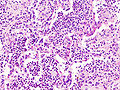
Aspiration pneumonia - 1 - (WC)
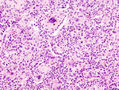
Aspiration pneumonia - 2 - (WC)
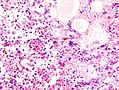
Aspiration pneumonia - 3 - (WC)
.jpg)
.jpg)
.jpg)
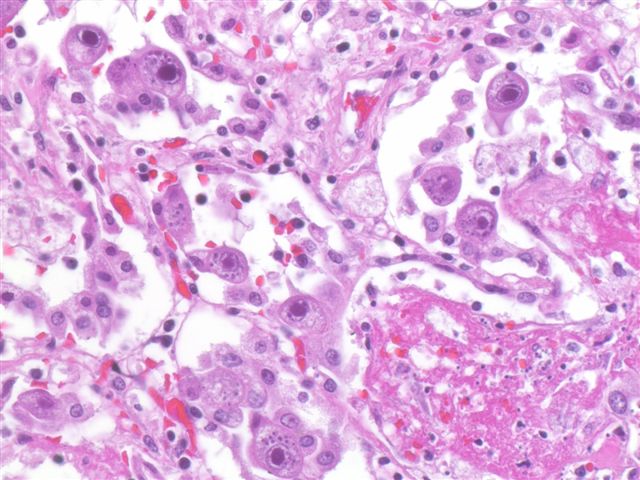
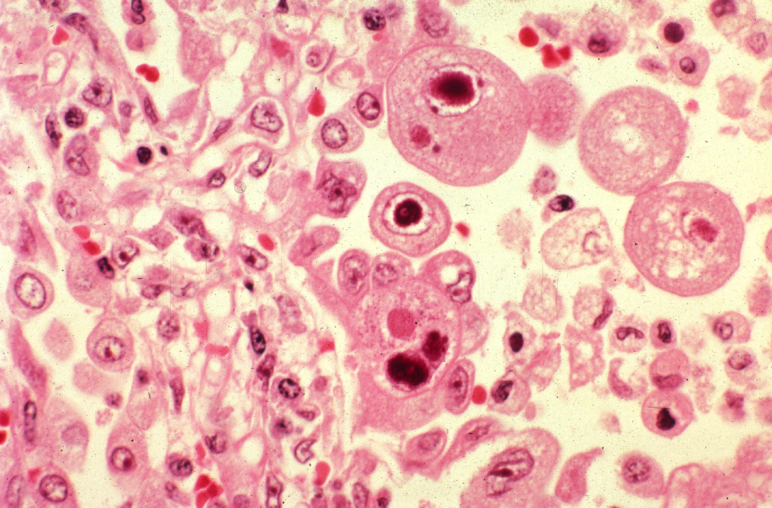
Cytomegalovirus pneumonia
Main article: Cytomegalovirus
General
- Immunodeficiency.
- Critical illness.[4]
Microscopic
Features:
- CMV nuclear changes:
- Large red nucleus with a pale halo.
- Eosinophilic granular cytoplasmic inclusions.
Images:
IHC
- CMV +ve -- cytoplasmic inclusions, large nucleus.
Diffuse lung diseases
Main article: Diffuse lung disease
- AKA idiopathic interstitial pneumonia.
Histologic pattern:
- Organizing pneumonia.
- Usual interstitial pneumonia.
- Nonspecific interstitial pneumonia.
- Lymphocytic interstitial pneumonia.
- Desquamative interstitial pneumonia.
- Diffuse alveolar damage.
See also
References
- ↑ Kumar, Vinay; Abbas, Abul K.; Fausto, Nelson; Aster, Jon (2009). Robbins and Cotran pathologic basis of disease (8th ed.). Elsevier Saunders. pp. 711. ISBN 978-1416031215.
- ↑ 2.0 2.1 Raghavendran, K.; Nemzek, J.; Napolitano, LM.; Knight, PR. (Apr 2011). "Aspiration-induced lung injury.". Crit Care Med 39 (4): 818-26. doi:10.1097/CCM.0b013e31820a856b. PMID 21263315.
- ↑ 3.0 3.1 Ohrui, T. (Sep 2005). "Preventive strategies for aspiration pneumonia in elderly disabled persons.". Tohoku J Exp Med 207 (1): 3-12. PMID 16082150.
- ↑ Limaye, AP.; Boeckh, M. (Nov 2010). "CMV in critically ill patients: pathogen or bystander?". Rev Med Virol 20 (6): 372-9. doi:10.1002/rmv.664. PMID 20931610.
- ↑ URL: http://www.pathologyoutlines.com/topic/lungnontumorCMV.html. Accessed on: 23 January 2012.