Pneumocytoma
Jump to navigation
Jump to search
The printable version is no longer supported and may have rendering errors. Please update your browser bookmarks and please use the default browser print function instead.
| Pneumocytoma | |
|---|---|
| Diagnosis in short | |
 Pneumocytoma. H&E stain. (WC/Nephron) | |
|
| |
| Synonyms | sclerosing hemangioma, benign pneumocytoma |
|
| |
| LM | mixed cell population, variable architecture (papillary, sclerotic, solid, hemorrhagic), +/-granulomas |
| LM DDx | carcinoid tumour, papillary pattern lung adenocarcinoma, metastatic papillary thyroid carcinoma |
| IHC | TTF-1 +ve, Ki-67 +ve membranous pattern, PR +ve, CD56 -ve, CD34 -ve |
| Site | lung - see lung tumours |
|
| |
| Clinical history | typical patient - female 40s |
| Prevalence | rare <= 1% of lung tumours |
| Radiology | slow growth/no growth, typically peripheral location |
| Prognosis | benign, case reports of mets |
| Clin. DDx | other lung tumours |
| Treatment | usually excision, may be followed |
Pneumocytoma is a rare lung tumour that is typically benign. It is also known as sclerosing pneumocytoma.[1][2]
It was previously known as sclerosing hemangioma.[2]
General
- Derived from type 2 pneumocyte.[3]
- Progesterone-receptor positive stromal cells.[4]
- Rare - 0.2% to 1% of lung tumours.[5]
- One large series had 100 cases.[6]
Management:
- Surgical excision preferred, may be followed.[5]
Epidemiology
- Female in 40s.[7]
- Considered benign; excision is curative.
- Rare case reports of metastases.[8][9]
Gross
- Peripheral, solitary.
- Well-circumscribed.
- Classically hemorrhagic.
Microscopic
Features:[7]
- Mixed cell population.
- Variable architecture:
- Papillary.
- Sclerotic.
- Solid.
- Hemorrhagic.
- +/-Granulomas.
DDx:[10]
- Papillary pattern:
- Papillary pattern lung adenocarcinoma.
- Metastatic papillary thyroid carcinoma.
- Solid pattern:
- Neuroendocrine tumour (carcinoid).
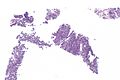
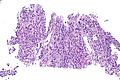
Images
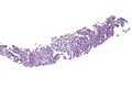
Pneumocytoma - low mag.
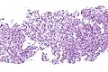
Pneumocytoma - intermed. mag.
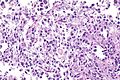
Pneumocytoma - high mag.
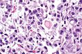
Pneumocytoma - very high mag.
Pneumocytoma - low mag.
Pneumocytoma - intermed. mag.


IHC
Features:[11]
Negative stains:[11]
- SMA -ve.
- CEA -ve.
- CD34 -ve.
- S100 -ve.
- Chromogranin A -ve.
Others:[3]
- TTF-1 +ve.
- HNF-3 alpha +ve.
- HNF-3 beta +ve.
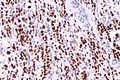
Images
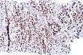
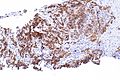
Pneumocytoma - TTF-1 - intermed. mag.
Pneumocytoma - TTF-1 - high mag.
Pneumocytoma - Ki-67 (membranous) - intermed. mag.
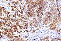
Pneumocytoma - Ki-67 (membranous) - high mag.


Sign out
Lung, Left Lower Lobe, Core Biopsy:
     - Sclerosing pneumocytoma (sclerosing hemangioma).

Comment:
The lesion stains as follows:
POSITIVE: Ki-67 (membranous pattern), TTF-1, PR.
NEGATIVE: CD56, p53, CD34.
Micro
The sections show lung with thickened alveolar walls containing bland appearing cells with round/oval nuclei without conspicuous nucleoli. Necrosis is absent.
See also
References
- ↑ Chan, KW.; Gibbs, AR.; Lo, WS.; Newman, GR. (Jun 1982). "Benign sclerosing pneumocytoma of lung (sclerosing haemangioma).". Thorax 37 (6): 404-12. PMID 6291188.
- ↑ 2.0 2.1 Ruiz de la Cuesta, D.; Lafont Rufat, M.; Ruiz de la Cuesta Martín, E. (Jun 2013). "Pneumocytoma (formerly known as sclerosing hemangioma of the lung): a rare cause of chest pain.". Arch Bronconeumol 49 (6): 276-7. doi:10.1016/j.arbres.2012.10.004. PMID 23380035.
- ↑ 3.0 3.1 Yamazaki, K. (Jul 2004). "Type-II pneumocyte differentiation in pulmonary sclerosing hemangioma: ultrastructural differentiation and immunohistochemical distribution of lineage-specific transcription factors (TTF-1, HNF-3 alpha, and HNF-3 beta) and surfactant proteins.". Virchows Arch 445 (1): 45-53. doi:10.1007/s00428-004-1023-3. PMID 15138814.
- ↑ 4.0 4.1 Einsfelder, BM.; Müller, KM. (Sep 2005). "["Pneumocytoma" or "sclerosing hemangioma": histogenetic aspects of a rare tumor of the lung]". Pathologe 26 (5): 367-77. doi:10.1007/s00292-005-0751-8. PMID 15731902.
- ↑ 5.0 5.1 Salemis, NS.; Seretis, C.; Nakos, G.; Kantounakis, I.; Stoumpos, C.; Spiliopoulos, K. (Jan 2013). "Synchronous occurrence of breast cancer and pulmonary sclerosing hemangioma: management and review of the literature.". Breast Dis 34 (2): 61-5. doi:10.3233/BD-130352. PMID 23838116.
- ↑ Devouassoux-Shisheboran, M.; Hayashi, T.; Linnoila, RI.; Koss, MN.; Travis, WD. (Jul 2000). "A clinicopathologic study of 100 cases of pulmonary sclerosing hemangioma with immunohistochemical studies: TTF-1 is expressed in both round and surface cells, suggesting an origin from primitive respiratory epithelium.". Am J Surg Pathol 24 (7): 906-16. PMID 10895813.
- ↑ 7.0 7.1 Keylock, JB.; Galvin, JR.; Franks, TJ. (May 2009). "Sclerosing hemangioma of the lung.". Arch Pathol Lab Med 133 (5): 820-5. PMID 19415961.
- ↑ Pokharel, S.; Dhillon, SS.; Ylagan, L.; George, S.; Yendamuri, S. (Oct 2016). "Sclerosing Pneumocytoma with Lymph Node Metastasis.". J Thorac Oncol 11 (10): 1802-4. doi:10.1016/j.jtho.2016.06.005. PMID 27346414.
- ↑ Tanaka, I.; Inoue, M.; Matsui, Y.; Oritsu, S.; Akiyama, O.; Takemura, T.; Fujiwara, M.; Kodama, T. et al. (Mar 1986). "A case of pneumocytoma (so-called sclerosing hemangioma) with lymph node metastasis.". Jpn J Clin Oncol 16 (1): 77-86. PMID 3009921.
- ↑ URL: http://www.med.muni.cz/biomedjournal/pdf/2004/01/37_42.pdf. Accessed on: 17 June 2010.
- ↑ 11.0 11.1 Rodriguez-Soto, J.; Colby, TV.; Rouse, RV. (Mar 2000). "A critical examination of the immunophenotype of pulmonary sclerosing hemangioma.". Am J Surg Pathol 24 (3): 442-50. PMID 10716159.
- ↑ Kim, BH.; Bae, YS.; Kim, SH.; Jeong, HJ.; Hong, SW.; Yoon, SO. (Feb 2013). "Usefulness of Ki-67 (MIB-1) immunostaining in the diagnosis of pulmonary sclerosing hemangiomas.". APMIS 121 (2): 105-10. doi:10.1111/j.1600-0463.2012.02945.x. PMID 23030396.