Difference between revisions of "Pneumocytoma"
Jump to navigation
Jump to search
| (4 intermediate revisions by the same user not shown) | |||
| Line 7: | Line 7: | ||
| Micro = mixed cell population, variable architecture (papillary, sclerotic, solid, hemorrhagic), +/-granulomas | | Micro = mixed cell population, variable architecture (papillary, sclerotic, solid, hemorrhagic), +/-granulomas | ||
| Subtypes = | | Subtypes = | ||
| LMDDx = [[carcinoid tumour]] | | LMDDx = [[carcinoid tumour]], papillary pattern [[lung adenocarcinoma]], metastatic [[papillary thyroid carcinoma]] | ||
| Stains = | | Stains = | ||
| IHC = TTF-1 +ve, Ki-67 +ve membranous pattern, PR +ve, CD56 -ve, CD34 -ve | | IHC = TTF-1 +ve, Ki-67 +ve membranous pattern, PR +ve, CD56 -ve, CD34 -ve | ||
| Line 38: | Line 38: | ||
*Progesterone-receptor positive stromal cells.<ref name=pmid15731902>{{Cite journal | last1 = Einsfelder | first1 = BM. | last2 = Müller | first2 = KM. | title = ["Pneumocytoma" or "sclerosing hemangioma": histogenetic aspects of a rare tumor of the lung] | journal = Pathologe | volume = 26 | issue = 5 | pages = 367-77 | month = Sep | year = 2005 | doi = 10.1007/s00292-005-0751-8 | PMID = 15731902 }}</ref> | *Progesterone-receptor positive stromal cells.<ref name=pmid15731902>{{Cite journal | last1 = Einsfelder | first1 = BM. | last2 = Müller | first2 = KM. | title = ["Pneumocytoma" or "sclerosing hemangioma": histogenetic aspects of a rare tumor of the lung] | journal = Pathologe | volume = 26 | issue = 5 | pages = 367-77 | month = Sep | year = 2005 | doi = 10.1007/s00292-005-0751-8 | PMID = 15731902 }}</ref> | ||
*Rare - 0.2% to 1% of lung tumours.<ref name=pmid23838116>{{Cite journal | last1 = Salemis | first1 = NS. | last2 = Seretis | first2 = C. | last3 = Nakos | first3 = G. | last4 = Kantounakis | first4 = I. | last5 = Stoumpos | first5 = C. | last6 = Spiliopoulos | first6 = K. | title = Synchronous occurrence of breast cancer and pulmonary sclerosing hemangioma: management and review of the literature. | journal = Breast Dis | volume = 34 | issue = 2 | pages = 61-5 | month = Jan | year = 2013 | doi = 10.3233/BD-130352 | PMID = 23838116 }}</ref> | *Rare - 0.2% to 1% of lung tumours.<ref name=pmid23838116>{{Cite journal | last1 = Salemis | first1 = NS. | last2 = Seretis | first2 = C. | last3 = Nakos | first3 = G. | last4 = Kantounakis | first4 = I. | last5 = Stoumpos | first5 = C. | last6 = Spiliopoulos | first6 = K. | title = Synchronous occurrence of breast cancer and pulmonary sclerosing hemangioma: management and review of the literature. | journal = Breast Dis | volume = 34 | issue = 2 | pages = 61-5 | month = Jan | year = 2013 | doi = 10.3233/BD-130352 | PMID = 23838116 }}</ref> | ||
**One large series had 100 cases.<ref name=pmid10895813>{{Cite journal | last1 = Devouassoux-Shisheboran | first1 = M. | last2 = Hayashi | first2 = T. | last3 = Linnoila | first3 = RI. | last4 = Koss | first4 = MN. | last5 = Travis | first5 = WD. | title = A clinicopathologic study of 100 cases of pulmonary sclerosing hemangioma with immunohistochemical studies: TTF-1 is expressed in both round and surface cells, suggesting an origin from primitive respiratory epithelium. | journal = Am J Surg Pathol | volume = 24 | issue = 7 | pages = 906-16 | month = Jul | year = 2000 | doi = | PMID = 10895813 }}</ref> | |||
Management: | Management: | ||
| Line 50: | Line 51: | ||
*Peripheral, solitary. | *Peripheral, solitary. | ||
*Well-circumscribed. | *Well-circumscribed. | ||
*Classically hemorrhagic. | |||
==Microscopic== | ==Microscopic== | ||
| Line 62: | Line 64: | ||
DDx:<ref>URL: [http://www.med.muni.cz/biomedjournal/pdf/2004/01/37_42.pdf http://www.med.muni.cz/biomedjournal/pdf/2004/01/37_42.pdf]. Accessed on: 17 June 2010.</ref> | DDx:<ref>URL: [http://www.med.muni.cz/biomedjournal/pdf/2004/01/37_42.pdf http://www.med.muni.cz/biomedjournal/pdf/2004/01/37_42.pdf]. Accessed on: 17 June 2010.</ref> | ||
*Papillary | *Papillary pattern: | ||
*[[Neuroendocrine tumour]] (carcinoid). | **Papillary pattern [[lung adenocarcinoma]]. | ||
**Metastatic [[papillary thyroid carcinoma]]. | |||
*Solid pattern: | |||
**[[Neuroendocrine tumour]] (carcinoid). | |||
===Images=== | ===Images=== | ||
| Line 114: | Line 119: | ||
===Micro=== | ===Micro=== | ||
The sections show lung with thickened alveolar walls containing bland appearing nuclei without conspicuous nucleoli. Necrosis is absent. | The sections show lung with thickened alveolar walls containing bland appearing cells with round/oval nuclei without conspicuous nucleoli. Necrosis is absent. | ||
==See also== | ==See also== | ||
Latest revision as of 23:23, 17 March 2019
| Pneumocytoma | |
|---|---|
| Diagnosis in short | |
 Pneumocytoma. H&E stain. (WC/Nephron) | |
|
| |
| Synonyms | sclerosing hemangioma, benign pneumocytoma |
|
| |
| LM | mixed cell population, variable architecture (papillary, sclerotic, solid, hemorrhagic), +/-granulomas |
| LM DDx | carcinoid tumour, papillary pattern lung adenocarcinoma, metastatic papillary thyroid carcinoma |
| IHC | TTF-1 +ve, Ki-67 +ve membranous pattern, PR +ve, CD56 -ve, CD34 -ve |
| Site | lung - see lung tumours |
|
| |
| Clinical history | typical patient - female 40s |
| Prevalence | rare <= 1% of lung tumours |
| Radiology | slow growth/no growth, typically peripheral location |
| Prognosis | benign, case reports of mets |
| Clin. DDx | other lung tumours |
| Treatment | usually excision, may be followed |
Pneumocytoma is a rare lung tumour that is typically benign. It is also known as sclerosing pneumocytoma.[1][2]
It was previously known as sclerosing hemangioma.[2]
General
- Derived from type 2 pneumocyte.[3]
- Progesterone-receptor positive stromal cells.[4]
- Rare - 0.2% to 1% of lung tumours.[5]
- One large series had 100 cases.[6]
Management:
- Surgical excision preferred, may be followed.[5]
Epidemiology
- Female in 40s.[7]
- Considered benign; excision is curative.
- Rare case reports of metastases.[8][9]
Gross
- Peripheral, solitary.
- Well-circumscribed.
- Classically hemorrhagic.
Microscopic
Features:[7]
- Mixed cell population.
- Variable architecture:
- Papillary.
- Sclerotic.
- Solid.
- Hemorrhagic.
- +/-Granulomas.
DDx:[10]
- Papillary pattern:
- Papillary pattern lung adenocarcinoma.
- Metastatic papillary thyroid carcinoma.
- Solid pattern:
- Neuroendocrine tumour (carcinoid).
Images
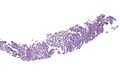
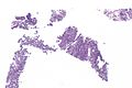
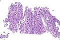
Pneumocytoma - low mag.
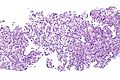
Pneumocytoma - intermed. mag.
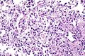
Pneumocytoma - high mag.
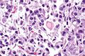
Pneumocytoma - very high mag.
Pneumocytoma - low mag.
Pneumocytoma - intermed. mag.


IHC
Features:[11]
Negative stains:[11]
- SMA -ve.
- CEA -ve.
- CD34 -ve.
- S100 -ve.
- Chromogranin A -ve.
Others:[3]
- TTF-1 +ve.
- HNF-3 alpha +ve.
- HNF-3 beta +ve.
Images
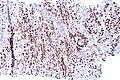
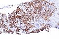
Pneumocytoma - TTF-1 - intermed. mag.
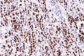
Pneumocytoma - TTF-1 - high mag.
Pneumocytoma - Ki-67 (membranous) - intermed. mag.
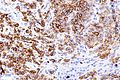
Pneumocytoma - Ki-67 (membranous) - high mag.


Sign out
Lung, Left Lower Lobe, Core Biopsy:
     - Sclerosing pneumocytoma (sclerosing hemangioma).

Comment:
The lesion stains as follows:
POSITIVE: Ki-67 (membranous pattern), TTF-1, PR.
NEGATIVE: CD56, p53, CD34.
Micro
The sections show lung with thickened alveolar walls containing bland appearing cells with round/oval nuclei without conspicuous nucleoli. Necrosis is absent.
See also
References
- ↑ Chan, KW.; Gibbs, AR.; Lo, WS.; Newman, GR. (Jun 1982). "Benign sclerosing pneumocytoma of lung (sclerosing haemangioma).". Thorax 37 (6): 404-12. PMID 6291188.
- ↑ 2.0 2.1 Ruiz de la Cuesta, D.; Lafont Rufat, M.; Ruiz de la Cuesta Martín, E. (Jun 2013). "Pneumocytoma (formerly known as sclerosing hemangioma of the lung): a rare cause of chest pain.". Arch Bronconeumol 49 (6): 276-7. doi:10.1016/j.arbres.2012.10.004. PMID 23380035.
- ↑ 3.0 3.1 Yamazaki, K. (Jul 2004). "Type-II pneumocyte differentiation in pulmonary sclerosing hemangioma: ultrastructural differentiation and immunohistochemical distribution of lineage-specific transcription factors (TTF-1, HNF-3 alpha, and HNF-3 beta) and surfactant proteins.". Virchows Arch 445 (1): 45-53. doi:10.1007/s00428-004-1023-3. PMID 15138814.
- ↑ 4.0 4.1 Einsfelder, BM.; Müller, KM. (Sep 2005). "["Pneumocytoma" or "sclerosing hemangioma": histogenetic aspects of a rare tumor of the lung]". Pathologe 26 (5): 367-77. doi:10.1007/s00292-005-0751-8. PMID 15731902.
- ↑ 5.0 5.1 Salemis, NS.; Seretis, C.; Nakos, G.; Kantounakis, I.; Stoumpos, C.; Spiliopoulos, K. (Jan 2013). "Synchronous occurrence of breast cancer and pulmonary sclerosing hemangioma: management and review of the literature.". Breast Dis 34 (2): 61-5. doi:10.3233/BD-130352. PMID 23838116.
- ↑ Devouassoux-Shisheboran, M.; Hayashi, T.; Linnoila, RI.; Koss, MN.; Travis, WD. (Jul 2000). "A clinicopathologic study of 100 cases of pulmonary sclerosing hemangioma with immunohistochemical studies: TTF-1 is expressed in both round and surface cells, suggesting an origin from primitive respiratory epithelium.". Am J Surg Pathol 24 (7): 906-16. PMID 10895813.
- ↑ 7.0 7.1 Keylock, JB.; Galvin, JR.; Franks, TJ. (May 2009). "Sclerosing hemangioma of the lung.". Arch Pathol Lab Med 133 (5): 820-5. PMID 19415961.
- ↑ Pokharel, S.; Dhillon, SS.; Ylagan, L.; George, S.; Yendamuri, S. (Oct 2016). "Sclerosing Pneumocytoma with Lymph Node Metastasis.". J Thorac Oncol 11 (10): 1802-4. doi:10.1016/j.jtho.2016.06.005. PMID 27346414.
- ↑ Tanaka, I.; Inoue, M.; Matsui, Y.; Oritsu, S.; Akiyama, O.; Takemura, T.; Fujiwara, M.; Kodama, T. et al. (Mar 1986). "A case of pneumocytoma (so-called sclerosing hemangioma) with lymph node metastasis.". Jpn J Clin Oncol 16 (1): 77-86. PMID 3009921.
- ↑ URL: http://www.med.muni.cz/biomedjournal/pdf/2004/01/37_42.pdf. Accessed on: 17 June 2010.
- ↑ 11.0 11.1 Rodriguez-Soto, J.; Colby, TV.; Rouse, RV. (Mar 2000). "A critical examination of the immunophenotype of pulmonary sclerosing hemangioma.". Am J Surg Pathol 24 (3): 442-50. PMID 10716159.
- ↑ Kim, BH.; Bae, YS.; Kim, SH.; Jeong, HJ.; Hong, SW.; Yoon, SO. (Feb 2013). "Usefulness of Ki-67 (MIB-1) immunostaining in the diagnosis of pulmonary sclerosing hemangiomas.". APMIS 121 (2): 105-10. doi:10.1111/j.1600-0463.2012.02945.x. PMID 23030396.