Pleural invasion

Pleural invasion is the presence of tumour in the pleura. Pleural invasion is considered a poor prognosticator in lung cancer,[1][2] and important in determining the tumour stage in lung cancer staging.
General

The visceral pleura is the pleura that covers the lung; tumour invasion into it is known as visceral pleural invasion (abbreviated VPI). The parietal pleural covers the chest wall.
The visceral pleura has two elastic layers:[3]
- External elastic layer.
- Usually thick/prominent.
- Important for staging.
- Internal elastic layer.
- Thin - may be difficult to see.
- Irrelevant for staging.
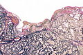
Images - normal pleural
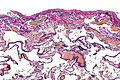
Lung visceral pleura - intermed. mag.
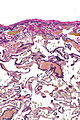
Lung visceral pleura - high mag.


Modified Hammar classification
Pleural invasion can be subgrouped as follows:[1]
| Category | Description | Tumour stage (TNM staging) |
|---|---|---|
| PL0 † | tumour confined to lung, not through elastic layer | T(any) |
| PL1 † | tumour beyond (external) elastic layer of the visercal pleura, not at pleural surface | at least T2 ‡ |
| PL2 | tumour at surface of pleura, not in parietal pleura | at least T2 |
| PL3 | tumour within parietal pleura and visceral pleura | at least T3 |
Notes:
- † The interface between PL0 and PL1 is the most challenging. Use of an elastic stain is recommend when there is uncertainty.[3]
- ‡ The up-staging of small tumours is not completely without controversy; there is data for lung adenocarcinomas less than 2 cm that suggests VPI is not an outcome predictor.[4]
Gross
- Tumour close to visceral pleura, e.g. <1 mm - high probability of VPI.
- Puckering of the visceral pleura overlying the tumour - suspicious for VPI.
Microscopic
Features - for PL1:
- Must be through the external elastic layer.[3]
Note:
- Tumour trapped within the (external) elastic layer is still PL0.[3]
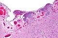
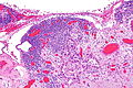
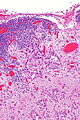
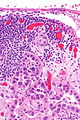
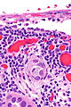
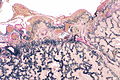
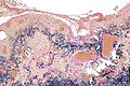
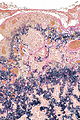
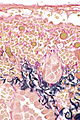
Images
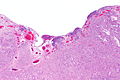
Adenocarcinoma - VPI - very low mag.
Adenocarcinoma - VPI - low mag.
Adenocarcinoma - VPI - intermed. mag.
Adenocarcinoma - VPI - intermed. mag.
Adenocarcinoma - VPI - high mag.
Adenocarcinoma - VPI - very high mag.


Stains

- Elastic stain (e.g. Miller, elastic trichrome, Verhoeff-van Gieson) - demonstrating tumour superficial to the (external) elastica.
Images
Adenocarcinoma - VPI - Miller - very low mag.
Adenocarcinoma - VPI - Miller - low mag.
Adenocarcinoma - VPI - Miller - intermed. mag.
Adenocarcinoma - VPI - Miller - intermed. mag.
Adenocarcinoma - VPI - Miller - very high mag.


See also
References
- ↑ 1.0 1.1 Travis, WD.; Brambilla, E.; Rami-Porta, R.; Vallières, E.; Tsuboi, M.; Rusch, V.; Goldstraw, P. (Dec 2008). "Visceral pleural invasion: pathologic criteria and use of elastic stains: proposal for the 7th edition of the TNM classification for lung cancer.". J Thorac Oncol 3 (12): 1384-90. doi:10.1097/JTO.0b013e31818e0d9f. PMID 19057261.
- ↑ Shimizu, K.; Yoshida, J.; Nagai, K.; Nishimura, M.; Ishii, G.; Morishita, Y.; Nishiwaki, Y. (Jul 2005). "Visceral pleural invasion is an invasive and aggressive indicator of non-small cell lung cancer.". J Thorac Cardiovasc Surg 130 (1): 160-5. doi:10.1016/j.jtcvs.2004.11.021. PMID 15999057.
- ↑ 3.0 3.1 3.2 3.3 Dacic, S. (Oct 2012). "Dilemmas in lung cancer staging.". Arch Pathol Lab Med 136 (10): 1194-7. doi:10.5858/arpa.2012-0282-CC. PMID 23020722.
- ↑ Nitadori, J.; Colovos, C.; Kadota, K.; Sima, CS.; Sarkaria, IS.; Rizk, NP.; Rusch, VW.; Travis, WD. et al. (Nov 2013). "Visceral pleural invasion does not affect recurrence or overall survival among patients with lung adenocarcinoma ≤ 2 cm: a proposal to reclassify T1 lung adenocarcinoma.". Chest 144 (5): 1622-31. doi:10.1378/chest.13-0394. PMID 23807749.