Placental infarct
Jump to navigation
Jump to search
| Placental infarct | |
|---|---|
| Diagnosis in short | |
 Placental infarct. H&E stain. | |
|
| |
| LM | necrosis of villi (hyaline material replaces the stroma of the villi) with loss of intervillous space, increased numbers of villi with syncytial knots (adjacent to infarct), thickened trophoblastic basement membrane (below cytotrophoblasts), +/-changes seen in decidual vasculopathy |
| LM DDx | perivillous fibrin deposition |
| Gross | lesion - red (early), white/grey (late) |
| Site | placenta |
|
| |
| Associated Dx | +/-hypertension |
| Prognosis | dependent on size and location |
Placental infarct is the death of placental tissue due to a compromised blood supply.
It should be noted that a maternal floor infarct is not a true infarct,[1] and is dealt with separately in its own article.
Placental ischemia can be thought of as a precursor to infarction; it redirects to this article.
General
- May be seen in conjunction with a retroplacental hematoma.
- Infarcts frequently associated with hypertension.[2][3]
- May be seen in fetal thrombotic vasculopathy.
Gross
Features:[4]
- Early - red.
- Late - white/grey.
Significant infarcts
- > 3cm --or-- central location --or-- in 1st or 2nd trimester.[citation needed]
- Small foci are accepted in term placentae - typically at periphery.
Images:
Microscopic
Features:
- Necrosis of villi; hyaline material (acellular eosinophilic material) replaces the stroma of the villi.
- Loss of intervillous space.[4]
- Villi appear to be crowded.[5]
- Normal spacing is ~1x smallest villus or larger.
- In perivillous fibrin deposition - spacing usu. larger than normal.
- Normal spacing is ~1x smallest villus or larger.
- Villi appear to be crowded.[5]
- Increased numbers of villi with syncytial knots. ‡
- Thickened trophoblastic basement membrane (below cytotrophoblasts).
- +/-Changes seen in decidual vasculopathy:
- Acute atherosis (vaguely like atherosclerosis).
- Fibrinoid necrosis.
- Vessel wall lipid deposition.
- Acute atherosis (vaguely like atherosclerosis).
Note:
- ‡ The normal number is dependent on the gestational age:[6]
| Gestational age | Villi with knots (average) |
Villi with knots (range) |
|---|---|---|
| 40 weeks | 29 % | 27-36 % |
| 39 weeks | 29 % | 23-34% |
| 38 weeks | 27 % | 23-33 % |
| 37 weeks | 28 % | 24-33 % |
| 36 weeks | 23 % | 20-26 % |
| 35 weeks | 22 % | 15-32 % |
| 34 weeks | 18 % | 12-22 % |
| 32 weeks | 13 % | 10-16 % |
| 30 weeks | 13 % | 10-17 % |
| 26 weeks | 11 % | 10-13 % |
| 22 weeks | 7 % | 5-9 % |
Images
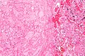
Placental infarct - low mag. (WC)

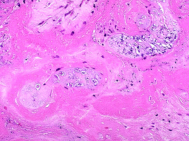
Placental infarct - intermed. mag. (WC)

www:
- Recent infarct (pathweb.uchc.edu).
- Placental infarct (umpmc.edu).[7]
- Placental infarct - necrotic villi (mda-sy.com).
Sign out
PLACENTA, UMBILICAL CORD AND FETAL MEMBRANES, BIRTH:
- THREE VESSEL UMBILICAL CORD WITHIN NORMAL LIMITS.
- FETAL MEMBRANES WITHIN NORMAL LIMITS.
- PLACENTAL DISC WITH THIRD TRIMESTER VILLI AND TWO PLACENTAL INFARCTS (0.6 CM AND
  0.4 CM IN MAXIMAL DIMENSION).
Increased syncytial knots
PLACENTA, UMBILICAL CORD AND FETAL MEMBRANES, BIRTH:
- THREE VESSEL UMBILICAL CORD WITHIN NORMAL LIMITS.
- FETAL MEMBRANES WITHIN NORMAL LIMITS.
- PLACENTAL DISC WITH THIRD TRIMESTER VILLI WITH FOCALLY INCREASED SYNCYTIAL KNOTS,
  AND MILD PERIVILLOUS FIBRIN DEPOSITION, OTHERWISE WITHIN NORMAL LIMITS.
See also
References
- ↑ Weedman Molavi, Diana (2008). The Practice of Surgical Pathology: A Beginner's Guide to the Diagnostic Process (1st ed.). Springer. pp. 178. ISBN 978-0387744858.
- ↑ URL: http://www.medind.nic.in/jae/t04/i1/jaet04i1p27.pdf. Accessed on: 16 April 2012.
- ↑ Becroft, DM.; Thompson, JM.; Mitchell, EA. (Apr 2002). "The epidemiology of placental infarction at term.". Placenta 23 (4): 343-51. doi:10.1053/plac.2001.0777. PMID 11969346.
- ↑ 4.0 4.1 Humphrey, Peter A; Dehner, Louis P; Pfeifer, John D (2008). The Washington Manual of Surgical Pathology (1st ed.). Lippincott Williams & Wilkins. pp. 465. ISBN 978-0781765275.
- ↑ Cotran, Ramzi S.; Kumar, Vinay; Fausto, Nelson; Nelso Fausto; Robbins, Stanley L.; Abbas, Abul K. (2005). Robbins and Cotran pathologic basis of disease (7th ed.). St. Louis, Mo: Elsevier Saunders. pp. 1109. ISBN 0-7216-0187-1.
- ↑ Loukeris, K.; Sela, R.; Baergen, RN.. "Syncytial knots as a reflection of placental maturity: reference values for 20 to 40 weeks' gestational age.". Pediatr Dev Pathol 13 (4): 305-9. doi:10.2350/09-08-0692-OA.1. PMID 20017638.
- ↑ URL: http://path.upmc.edu/cases/case75/micro.html. Accessed on: 6 January 2011.