Difference between revisions of "Perineurioma"
Jump to navigation
Jump to search
Jensflorian (talk | contribs) (molecular) |
Jensflorian (talk | contribs) (→Macroscopy: +Sclerosing perineurioma) |
||
| Line 13: | Line 13: | ||
*Intraneural perineurioma: segmental tubular enlargement of the nerve. | *Intraneural perineurioma: segmental tubular enlargement of the nerve. | ||
*Soft tissue perineurioma: Well circumscribed, but not encapsulated. | *Soft tissue perineurioma: Well circumscribed, but not encapsulated. | ||
*Sclerosing perineurioma: Predilection for hands.<ref name="pmid9414186">{{cite journal |vauthors=Fetsch JF, Miettinen M |title=Sclerosing perineurioma: a clinicopathologic study of 19 cases of a distinctive soft tissue lesion with a predilection for the fingers and palms of young adults |journal=Am. J. Surg. Pathol. |volume=21 |issue=12 |pages=1433–42 |date=December 1997 |pmid=9414186 |doi=10.1097/00000478-199712000-00005 |url=}}</ref> | |||
==Microscopic== | ==Microscopic== | ||
Revision as of 08:28, 10 September 2020
Perineurioma is an uncommon benign peripheral nerve sheath tumour.
General
- Benign tumour derived from perineurial cells (intraneural or soft tissue).
- ICD-O 9571/0
- WHO grade I
- Rarely malignant soft tissue perineurioma.
Variant:
- Reticular perineurioma.[1]
Macroscopy
- Intraneural perineurioma: segmental tubular enlargement of the nerve.
- Soft tissue perineurioma: Well circumscribed, but not encapsulated.
- Sclerosing perineurioma: Predilection for hands.[2]
Microscopic
Features:[3]
- Perineural epithelioid cells.
- Abundant pale, fluffy appearing cytoplasm.
Note:
- May be intraneural.[3]
- Typical pseudo-onion bulbs.
- Long considered hypertrophic neuropathy.
- Rare (less than 1% of all nerve sheath tumours).
DDx:
- Neuroma.
- Neurofibroma.
- Schwannoma.
- S100 +ve, EMA -ve.[3]
- Liposarcoma - reticular perineurioma.
Images:
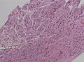
Pseudo-onion bulbs in a intraneural perineurioma (WC/jensflorian)

IHC
- S100 -ve.
- EMA +ve.
- CD34 ~65% +ve.[4]
Molecular
- Frequent TRAF7 mut.[6]
See also
References
- ↑ Graadt van Roggen, JF.; McMenamin, ME.; Belchis, DA.; Nielsen, GP.; Rosenberg, AE.; Fletcher, CD. (Apr 2001). "Reticular perineurioma: a distinctive variant of soft tissue perineurioma.". Am J Surg Pathol 25 (4): 485-93. PMID 11257623.
- ↑ "Sclerosing perineurioma: a clinicopathologic study of 19 cases of a distinctive soft tissue lesion with a predilection for the fingers and palms of young adults". Am. J. Surg. Pathol. 21 (12): 1433–42. December 1997. doi:10.1097/00000478-199712000-00005. PMID 9414186.
- ↑ 3.0 3.1 3.2 Mills, Stacey E; Carter, Darryl; Greenson, Joel K; Reuter, Victor E; Stoler, Mark H (2009). Sternberg's Diagnostic Surgical Pathology (5th ed.). Lippincott Williams & Wilkins. pp. 424. ISBN 978-0781779425.
- ↑ 4.0 4.1 Hornick, JL.; Fletcher, CD. (Jul 2005). "Soft tissue perineurioma: clinicopathologic analysis of 81 cases including those with atypical histologic features.". Am J Surg Pathol 29 (7): 845-58. PMID 15958848.
- ↑ Tsang, WY.; Chan, JK.; Chow, LT.; Tse, CC. (Aug 1992). "Perineurioma: an uncommon soft tissue neoplasm distinct from localized hypertrophic neuropathy and neurofibroma.". Am J Surg Pathol 16 (8): 756-63. PMID 1497116.
- ↑ "Genomic analysis reveals frequent TRAF7 mutations in intraneural perineuriomas". Ann. Neurol. 81 (2): 316–321. February 2017. doi:10.1002/ana.24854. PMC 5725965. PMID 28019650. https://www.ncbi.nlm.nih.gov/pmc/articles/PMC5725965/.