Difference between revisions of "Paneth cell"
Jump to navigation
Jump to search

(+images) |
|||
| Line 11: | Line 11: | ||
If PCM is present: | If PCM is present: | ||
*Think of [[inflammatory bowel disease]] and other long-standing injurious processes. | *Think of [[inflammatory bowel disease]] and other long-standing injurious processes. | ||
==Microscopic== | ==Microscopic== | ||
| Line 32: | Line 29: | ||
Image: Paneth cell metaplasia in the left colon -- extremely high mag.jpg | PCM - extremely high mag. (WC) | Image: Paneth cell metaplasia in the left colon -- extremely high mag.jpg | PCM - extremely high mag. (WC) | ||
</gallery> | </gallery> | ||
==IHC== | |||
*Lysozyme +ve.<ref name=pmid12655793>{{cite journal |author=Rubio CA, Nesi G |title=A simple method to demonstrate normal and metaplastic Paneth cells in tissue sections |journal=In Vivo |volume=17 |issue=1 |pages=67–71 |year=2003 |pmid=12655793 |doi= |url=}}</ref> | |||
==See also== | ==See also== | ||
Revision as of 11:43, 20 January 2014

Paneth cell metaplasia. H&E stain.
The Paneth cell is characteristic of the small intestine. It is also normal in the cecum, ascending colon and transverse colon.
Paneth cell metaplasia, abbreviated PCM, redirects to this article.
General
- Paneth cells should not be in the left colon.[1]
- If you see 'em there it is Paneth cell metaplasia.
Paneth cell metaplasia
If PCM is present:
- Think of inflammatory bowel disease and other long-standing injurious processes.
Microscopic
Features:
- Supranuclear eosinophilic granules.
DDx:
- Enterochromaffin cells (AKA Kulchitsky cells).
- Subnuclear eosinophilic granules.
- Intraepithelial eosinophils.
- Eosinophils have smaller (~1/2) more intensely red granules.
Images
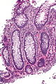
PCM - intermed. mag. (WC)
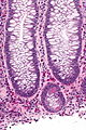
PCM - high mag. (WC)
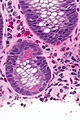
PCM - very high mag. (WC)
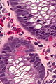
PCM - extremely high mag. (WC)


IHC
- Lysozyme +ve.[2]
See also
References
- ↑ Tanaka M, Saito H, Kusumi T, et al (December 2001). "Spatial distribution and histogenesis of colorectal Paneth cell metaplasia in idiopathic inflammatory bowel disease". J. Gastroenterol. Hepatol. 16 (12): 1353–9. PMID 11851832. http://www3.interscience.wiley.com/resolve/openurl?genre=article&sid=nlm:pubmed&issn=0815-9319&date=2001&volume=16&issue=12&spage=1353.
- ↑ Rubio CA, Nesi G (2003). "A simple method to demonstrate normal and metaplastic Paneth cells in tissue sections". In Vivo 17 (1): 67–71. PMID 12655793.