PEComa
Jump to navigation
Jump to search
The printable version is no longer supported and may have rendering errors. Please update your browser bookmarks and please use the default browser print function instead.
PEComa is a family of tumours derived from perivascular epithelioid cells (PECs).
General
- Associated with abnormalities in TSC1 and TSC2 - the genes involved in tuberous sclerosis.[1]
The PEComa family
- Angiomyolipoma.
- Lymphangioleiomyomatosis.
- Clear-cell myomelanocytic tumour of ligamentum teres/falciform ligament.
- Abdominopelvic sarcoma of perivascular epitheloid cells.[2]
- Clear-cell sugar tumour (CCST).
- Primary extrapulmonary sugar tumour.
Microscopic
Features:[1]
- Epithelioid morphology.
- Clear or granular cytoplasm.
- Central oval (or round) nucleus.
- Indistinct/small nucleolus.
DDx:
- Clear cell sarcoma.
- Melanotic Xp11 translocation renal cancer - in kidney.
- Other clear cell tumours.
- Other large epithelioid tumours.
Images
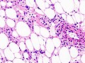
Renal AML (WC/KGH)
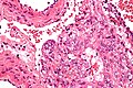
LAM - very high mag. (WC/Nephron)
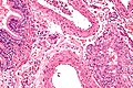
LAM - high mag. (WC/Nephron)
.jpg)


IHC
- Melanocytic markers
- Myogenic markers
- Calponin.
- Actin.[1]
- Myosin.
EM
- Premelanosomes.[4]
See also
References
- ↑ 1.0 1.1 1.2 1.3 Martignoni G, Pea M, Reghellin D, Zamboni G, Bonetti F (February 2008). "PEComas: the past, the present and the future". Virchows Arch. 452 (2): 119–32. doi:10.1007/s00428-007-0509-1. PMC 2234444. PMID 18080139. https://www.ncbi.nlm.nih.gov/pmc/articles/PMC2234444/.
- ↑ Bonetti, F.; Martignoni, G.; Colato, C.; Manfrin, E.; Gambacorta, M.; Faleri, M.; Bacchi, C.; Sin, VC. et al. (Jun 2001). "Abdominopelvic sarcoma of perivascular epithelioid cells. Report of four cases in young women, one with tuberous sclerosis.". Mod Pathol 14 (6): 563-8. doi:10.1038/modpathol.3880351. PMID 11406657.
- ↑ Doyle, LA.; Hornick, JL.; Fletcher, CD. (Dec 2013). "PEComa of the gastrointestinal tract: clinicopathologic study of 35 cases with evaluation of prognostic parameters.". Am J Surg Pathol 37 (12): 1769-82. doi:10.1097/PAS.0b013e31829caab3. PMID 24061520.
- ↑ Park, SH.; Ro, JY.; Kim, HS.; Lee, ES. (Nov 2003). "Perivascular epithelioid cell tumor of the uterus: immunohistochemical, ultrastructural and molecular study.". Pathol Int 53 (11): 800-5. PMID 14629307.