Ovary
Jump to navigation
Jump to search
The printable version is no longer supported and may have rendering errors. Please update your browser bookmarks and please use the default browser print function instead.
The ovary has a wealth of pathology. It has benign tumours and malignant ones. It is a significant part of gynecologic pathology.
Normal ovary
- Corpora albicans - pale/white body with lobulated contour.
- Involuted corpus luteum.
- Not seen pre-pubertal.
- Number increase with age.
- Ovarian follicles.
- Stroma - hyperchromatic - spindle morphology, whorling.
- If the cells have a round morphology... think about endometriosis.
Images
www:
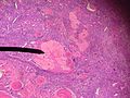
Corpus albicans. (WC)

Cysts - overview
General
- Very common.
Most common:
- Serous cystadenoma.
- Usually uniloculated.
- Morphology: ciliated, columnar.
- Mucinous cystadenoma.
- Usually multiloculated.[1]
- Memory device: multiloculated = mucinous.
- Usually multiloculated.[1]
- Endometrioma (see endometriosis).
- Simple cyst.
- Corpus luteum cyst.
- Cancerous cyst (see ovarian cancer).
Notes:
- Epithelium is often lost in processing - may make interpretation challenging
- Ovarian surface epithelium (previously call germinal epithelium) - covers the ovary
Ovarian surface vs. mesothelium:
- Image: ovarian surface epithelium - endojournals.org.
- Image: mesothelium - internetattitude.com.
Specific benign diagnoses
Endometriosis
Main article: Endometriosis
Corpus luteum cyst
General
- Normal in childbearing age women.
Gross
- Classically yellow.
Microscopic
Features:
- Pseudocyst lined by stratified, pale staining (luteinized) cells.
- +/-Hemorrhagic centre.
Images:
Benign mesothelial inclusion cyst
- AKA mesothelial inclusion cyst.
- AKA peritoneal inclusion cyst.[citation needed]
- AKA cortical inclusion cyst.[4][citation needed]
- AKA surface epithelial inclusion cyst.
General
- May be found incidentally, e.g. during C-section.
Epidemiology:
- Associated with previous surgery.
Gross
Microscopic
Features:
- Benign mesothelium.
- Single layer of squamoid or cuboid mesothelial cells.[6]
DDx:
- Serous cystadenoma of the ovary - must be >=1 cm.[7]
Image:
IHC
- CK +ve, calretinin +ve.[6]
Sign out
OVARY, LEFT, BIOPSY:
- BENIGN CORTICAL INCLUSION CYST.
Ovarian infarct
Main article: Ovarian infarct
Pregnancy luteoma
Main article: Pregnancy luteoma
Ovarian tumours
Main article: Ovarian tumours
For a very brief overview of gynecologic tumours see: Gynecologic pathology.
See also
References
- ↑ IAV. 6 February 2009.
- ↑ Auersperg N, Wong AS, Choi KC, Kang SK, Leung PC (April 2001). "Ovarian surface epithelium: biology, endocrinology, and pathology". Endocr. Rev. 22 (2): 255–88. PMID 11294827. http://edrv.endojournals.org/cgi/pmidlookup?view=long&pmid=11294827.
- ↑ ALS. 5 February 2009.
- ↑ Feeley, KM.; Wells, M. (Feb 2001). "Precursor lesions of ovarian epithelial malignancy.". Histopathology 38 (2): 87-95. PMID 11207821.
- ↑ GAG 26 Feb 2009.
- ↑ 6.0 6.1 6.2 Urbanczyk K, Skotniczny K, Kucinski J, Friediger J (2005). "Mesothelial inclusion cysts (so-called benign cystic mesothelioma)--a clinicopathological analysis of six cases". Pol J Pathol 56 (2): 81-7. PMID 16092670.
- ↑ Nucci, Marisa R.; Oliva, Esther (2009). Gynecologic Pathology: A Volume in Foundations in Diagnostic Pathology Series (1st ed.). Churchill Livingstone. pp. 384. ISBN 978-0443069208.
- ↑ Asch, E.; Levine, D.; Kim, Y.; Hecht, JL. (Mar 2008). "Histologic, surgical, and imaging correlations of adnexal masses.". J Ultrasound Med 27 (3): 327-42. PMID 18314510.