Ovarian fibroma
Jump to navigation
Jump to search
The printable version is no longer supported and may have rendering errors. Please update your browser bookmarks and please use the default browser print function instead.
| Ovarian fibroma | |
|---|---|
| Diagnosis in short | |
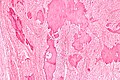
 Ovarian fibroma. H&E stain. | |
|
| |
| LM |
spindle cells with a central nucleus and no nuclear atypia; patternless pattern (not fascicular, not herring bone), +/-calcification |
| LM DDx | thecoma, leiomyoma, fibrosarcoma, metastatic metaplastic carcinoma, endometriosis with fibrosis |
| IHC | inhibin -ve |
| Gross | solid white mass, usu. well-circumscribed |
| Site | ovary - see ovarian tumours |
|
| |
| Syndromes | Meigs syndrome, nevoid basal cell carcinoma syndrome (esp. if calcified) |
|
| |
| Prevalence | uncommon |
| Prognosis | benign |
| Clin. DDx | other ovarian tumours - esp. solid ones |
Ovarian fibroma is a benign ovarian tumour.
General
- May be a part of:
- Meigs syndrome (mnemonic FAR: fibroma, ascites, right pleural effusion).
- Nevoid basal cell carcinoma syndrome (NBCCS), AKA Gorlin syndrome.[1]
- In NBCCS classically - calcified and bilateral.[2]
- Very rarely transform to fibrosarcoma <1%.[3]
Gross
Features:
- Solid white mass, usually well-circumscribed.[4]
Note:
- Thecoma = yellow solid mass.[4]
Images
www:
Microscopic
- Spindle cells with central nucleus and no nuclear atypia.
- Patternless pattern (AKA storiform pattern) - not fascicular, not herring bone.
- Stainable lipid - minimal or none.[6]
Notes:
- May be cellular.
- Mitotic activity minimal.[7]
DDx:
- Thecoma - lipid.
- Leiomyoma - fascicular architecture, rare in the ovary.[8]
- Fibrosarcoma - nuclear atypia, classically herring bone pattern, very rare.
- Metastatic metaplastic carcinoma - nuclear atypia, rare.
- Endometriosis with extensive fibrosis.
Images
Ovarian fibroma - intermed mag. (WC)
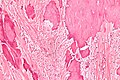
Ovarian fibroma - high mag. (WC)

IHC
- Inhibin -ve (~75%).[6]
Sign out
 OVARIAN MASS ("FIBROMA"), LEFT, EXCISION:
- FIBROMA.
- NEGATIVE FOR MALIGNANCY.
Micro
The sections show spindle cells in a patternless pattern. There is no appreciable nuclear atypia. No mitotic activity is apparent. No necrosis is identified. No calcifications are seen. A small amount of benign ovarian parenchyma is present at the edge of the lesion.
See also
References
- ↑ Cotran, Ramzi S.; Kumar, Vinay; Fausto, Nelson; Nelso Fausto; Robbins, Stanley L.; Abbas, Abul K. (2005). Robbins and Cotran pathologic basis of disease (7th ed.). St. Louis, Mo: Elsevier Saunders. pp. 1103. ISBN 0-7216-0187-1.
- ↑ Tytle, T.; Rosin, D. (Sep 1984). "Bilateral calcified ovarian fibromas.". South Med J 77 (9): 1178-80. PMID 6385289.
- ↑ URL: http://brighamrad.harvard.edu/Cases/bwh/hcache/353/full.html. Accessed on: 4 October 2011.
- ↑ 4.0 4.1 Rose, Alan G. (2008). Atlas of Gross Pathology with Histologic Correlation (1st ed.). Cambridge University Press. pp. 398. ISBN 978-0521868792.
- ↑ URL: http://www.pathologyoutlines.com/ovarytumor.html#fibroma. Accessed on: 7 May 2012.
- ↑ 6.0 6.1 6.2 Roth, LM. (Jul 2006). "Recent advances in the pathology and classification of ovarian sex cord-stromal tumors.". Int J Gynecol Pathol 25 (3): 199-215. doi:10.1097/01.pgp.0000192271.22289.e6. PMID 16810055.
- ↑ Huang, L.; Liao, LM.; Wang, HY.; Zheng, M. (2010). "Clinicopathologic characteristics and prognostic factors of ovarian fibrosarcoma: the results of a multi-center retrospective study.". BMC Cancer 10: 585. doi:10.1186/1471-2407-10-585. PMID 20979607.
- ↑ Rajabi, P.; Hani, M.; Bagheri, M.; Mirzadeh, F. (2014). "Large ovarian leiomyoma in young woman.". Adv Biomed Res 3: 88. doi:10.4103/2277-9175.128001. PMID 24761396.