Osteosarcoma
Jump to navigation
Jump to search
The printable version is no longer supported and may have rendering errors. Please update your browser bookmarks and please use the default browser print function instead.
| Osteosarcoma | |
|---|---|
| Diagnosis in short | |
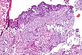
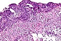
 Osteosarcoma. H&E stain. | |
|
| |
| LM | cells with malignant features (e.g. nuclear membrane irregularities, marked nuclear size differences, mitoses) surrounded by delicate strands of osteoid |
| Subtypes | conventional osteosarcoma (osteoblastic osteosarcoma, fibroblastic osteosarcoma, chondroblastic osteosarcoma), small cell osteosarcoma, telangiectatic osteosarcoma, parosteal osteosarcoma, periosteal osteosarcoma, low-grade central osteosarcoma, high-grade surface osteosarcoma, secondary osteosarcoma, gnathic osteosarcoma |
| LM DDx | chondrosarcoma, phosphaturic mesenchymal tumour, mixed connective tissue type, undifferentiated pleomorphic sarcoma (for fibroblastic osteosarcoma), aneurysmal bone cyst (for telangiectatic osteosarcoma), fibrous dysplasia, small round cell tumours (for small cell osteosarcoma) |
| Site | bone |
|
| |
| Syndromes | Li-Fraumeni syndrome |
|
| |
| Prevalence | uncommon |
| Clin. DDx | aneurysmal bone cyst,fibrous dysplasia, Ewing sarcoma |
Osteosarcoma, also known as osteogenic sarcoma, is a malignant bone tumour. It is grouped with the chondro-osseous tumours.
General
- Most common malignant bone tumour in children.
- May be seen in the context of Li-Fraumeni syndrome.
Trivia:
- Terry Fox was afflicited by this tumour.
Definition
- Tumour that makes osteoid.
- Osteoid = (extracellular) organic component of bone, normally produced by osteoblasts (cells which make bone matrix).
Gross
Classic locations:[1]
- Distal femur ~ 45%.
- Proximal tibia ~ 20%.
- Proximal humerous ~ 15%.
Microscopic
Features:
- Cells with malignant features (e.g. nuclear membrane irregularities, marked nuclear size differences, mitoses) surrounded by delicate strands of osteoid.
- Osteoid on H&E: pink, homogenous, "glassy".
- Tumours typically very cellular - when compared to normal bone.
- +/-Large (multinucleated) osteoclast-like giant cells.[2]
DDx:
Images
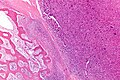
Osteosarcoma - low mag. (WC)
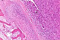
Osteosarcoma - intermed. mag. (WC)
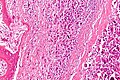
Osteosarcoma - high mag. (WC)
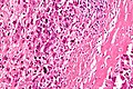
Osteosarcoma - very high mag. (WC)
Small cell osteosarcoma - intermed. mag. (WC)
Small cell osteosarcoma - high mag. (WC)


www:
Subtypes
- Conventional osteosarcoma (high grade).
- Osteoblastic osteosarcoma.
- Fibroblastic osteosarcoma.
- Chondroblastic osteosarcoma.
- Small cell osteosarcoma.
- Telangiectatic osteosarcoma.
- Parosteal osteosarcoma.
- Periosteal osteosarcoma.
- Low-grade central osteosarcoma.
- High-grade surface osteosarcoma.
- Secondary osteosarcoma.
- Gnathic osteosarcoma - jaw bones - usually chondroblastic.
How to remember:
- Convention FOC = fibroblastic, osteogenic, chondroblastic.
- Low-grade central.
- High-grade surface.
- Parosteal.
- Periosteal.
- Small cell.
- Secondary.
- Telangiectatic.
Chondroblastic osteosarcoma
- Chondroid matrix present - may be prominent; osteoid may be a minor component.
- May be confused with chondrosarcoma.
Fibroblastic osteosarcoma
- Undifferentiated pleomorphic sarcoma-like/MFH-like.
Images:
Low-grade central osteosarcoma
- Well-formed bone.
- Usu. minimal nuclear atypia.
DDx:
Telangiectatic osteosarcoma
Important radiologic DDx:
Parosteal osteosarcoma
DDx:
Periosteal osteosarcoma
- Intermediate grade.[10]
Small cell osteosarcoma
- May mimic (other) small round cell tumours.
Secondary osteosarcoma
Arises in the context of something else - causes:
- Paget disease of the bone (~80% of secondary osteosarcomas)
- Radiation (~15% of secondary osteosarcomas)).[11]
- Prognosis often poor.[10]
Images:
See also
References
- ↑ Greenwald, J.; Heng, M. (2007). Toronto Notes for Medical Students 2007 (2007 ed.). The Toronto Notes Inc. for Medical Students Inc.. pp. OR43. ISBN 978-0968592878.
- ↑ Papalas JA, Balmer NN, Wallace C, Sangueeza OP (June 2009). "Ossifying dermatofibroma with osteoclast-like giant cells: report of a case and literature review". Am J Dermatopathol 31 (4): 379-83. doi:10.1097/DAD.0b013e3181966747. PMID 19461244.
- ↑ Papandreou, C.; Skopelitou, A.; Kappes, G.; Daouaher, H. (2010). "Primary osteosarcoma of the urinary bladder treated with external radiotherapy in a patient with a history of transitional cell carcinoma: a case report.". J Med Case Rep 4: 70. doi:10.1186/1752-1947-4-70. PMC 2843711. PMID 20181254. https://www.ncbi.nlm.nih.gov/pmc/articles/PMC2843711/.
- ↑ Humphrey, Peter A; Dehner, Louis P; Pfeifer, John D (2008). The Washington Manual of Surgical Pathology (1st ed.). Lippincott Williams & Wilkins. pp. 638. ISBN 978-0781765275.
- ↑ URL: http://bestpractice.bmj.com/best-practice/monograph/780/basics/classification.html. Accessed on: 7 April 2011.
- ↑ Inwards, CY (2001). "Low-grade central osteosarcoma versus fibrous dysplasia". Pathology Case Reviews 6 (1): 22-27. http://journals.lww.com/pathologycasereviews/Fulltext/2001/01000/Low_Grade_Central_Osteosarcoma_Versus_Fibrous.5.aspx.
- ↑ Patibandla, MR.; Uppin, SG.; Thotakura, AK.; Panigrahi, MK.; Challa, S.. "Primary telangiectatic osteosarcoma of occipital bone: a case report and review of literature.". Neurol India 59 (1): 117-9. doi:10.4103/0028-3886.76891. PMID 21339678.
- ↑ Weiss, A.; Khoury, JD.; Hoffer, FA.; Wu, J.; Billups, CA.; Heck, RK.; Quintana, J.; Poe, D. et al. (Apr 2007). "Telangiectatic osteosarcoma: the St. Jude Children's Research Hospital's experience.". Cancer 109 (8): 1627-37. doi:10.1002/cncr.22574. PMID 17351949.
- ↑ The International Agency for Research on Cancer (Editors: Fletcher, C.D.M.; Unni, K. Krishnan; Mertens, F.) (2006). Pathology and Genetics of Tumours of Soft Tissue and Bone (IARC WHO Classification of Tumours) (3rd ed.). World Health Organization. pp. 279. ISBN 978-9283224136.
- ↑ 10.0 10.1 10.2 Carrle, D.; Bielack, SS. (Dec 2006). "Current strategies of chemotherapy in osteosarcoma.". Int Orthop 30 (6): 445-51. doi:10.1007/s00264-006-0192-x. PMC 3172747. PMID 16896870. https://www.ncbi.nlm.nih.gov/pmc/articles/PMC3172747/.
- ↑ URL: http://www.rsna.org/REG/publications/rg/afip/privateM/1997/0017/0005/1205/6.htm. Accessed on: 8 April 2011.