Osteoblastoma
Jump to navigation
Jump to search
| Osteoblastoma | |
|---|---|
| Diagnosis in short | |
 Osteoblastoma. H&E stain. | |
|
| |
| LM | anastomosing bony trabeculae with variable mineralization, osteoblast rimming, no nuclear atypia of osteocytes |
| LM DDx | osteoid osteoma, osteosarcoma |
| Radiology | > 1.5 cm (smaller lesions osteoid osteoma) |
| Clin. DDx | osteosarcoma |
Osteoblastoma is benign primary bone tumour.
General
- Benign bone tumour.
- Uncommon.[1]
Gross
Microscopic
Features:[4]
- Anastomosing bony trabeculae with:
- Osteoblasts rimming.
- Cells line-up at edge of bone.
- Osteoblasts rimming.
Notes:
- Histomorphologically near identical/indistinguishable from osteoid osteoma.[3]
Images
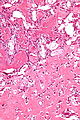
Osteoblastoma - high mag. (WC)
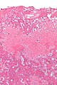
Osteoblastoma - low mag. (WC)

Sign out
BONE, LEFT FEMUR, EXCISION:
- OSTEOBLASTOMA.
See also
References
- ↑ Khan, IS.; Thakur, JD.; Chittiboina, P.; Nanda, A.. "Large sacral osteoblastoma: a case report and review of multi-disciplinary management strategies.". J La State Med Soc 164 (5): 251-5. PMID 23362588.
- ↑ Boriani, S.; Amendola, L.; Bandiera, S.; Simoes, CE.; Alberghini, M.; Di Fiore, M.; Gasbarrini, A. (Oct 2012). "Staging and treatment of osteoblastoma in the mobile spine: a review of 51 cases.". Eur Spine J 21 (10): 2003-10. doi:10.1007/s00586-012-2395-8. PMID 22695702.
- ↑ 3.0 3.1 Mills, Stacey E; Carter, Darryl; Greenson, Joel K; Oberman, Harold A; Reuter, Victor E (2004). Sternberg's Diagnostic Surgical Pathology (4th ed.). Lippincott Williams & Wilkins. pp. 286. ISBN 978-0781740517.
- ↑ Mills, Stacey E; Carter, Darryl; Greenson, Joel K; Oberman, Harold A; Reuter, Victor E (2004). Sternberg's Diagnostic Surgical Pathology (4th ed.). Lippincott Williams & Wilkins. pp. 285. ISBN 978-0781740517.