Organizing pneumonia
Jump to navigation
Jump to search
The printable version is no longer supported and may have rendering errors. Please update your browser bookmarks and please use the default browser print function instead.
| Organizing pneumonia | |
|---|---|
| Diagnosis in short | |
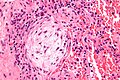
 Organizing pneumonia. H&E stain. | |
|
| |
| LM | distal airway disease -- airways plugged with organizing exudate (fluffy light-staining paucicellular regions with stellate cells); no hobnailing of pneumocytes; type 2 pneumocytes hyperplasia is absent |
| LM DDx | diffuse alveolar damage (proliferative phase), bronchiolitis obliterans. |
| Site | lung - diffuse lung diseases |
|
| |
| Prevalence | uncommon |
| Prognosis | dependent on underlying cause |
| Clin. DDx | cryptogenic organizing pneumonia, transplant rejection, infection (pneumonia), collagen vascular disease, peri-tumour |
| Treatment | dependent on underlying cause |
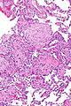
Organizing pneumonia, abbreviated OP, is a histologic pattern in lung pathology. It fits into the larger category of diffuse lung diseases.
General
- Multiple causes, e.g. transplant rejection, infection.
Clinical diagnoses:[1]
- Transplant rejection.
- Cryptogenic organizing pneumonia (COP), AKA (idiopathic) bronchiolitis obliterans organizing pneumonia (BOOP).
- Should not be confused with constrictive bronchiolitis (AKA bronchiolitis obliterans).
- Collagen vascular disease.
- Toxic injury.
- Infection.
- Peri-tumour - in proximity to a space-occupying lesion (abscess, neoplasm).
Note:
- BOOP is used as a synonym for organizing pneumonia which has the long differential diagnosis above.[1]
- Confusingly, it may be used to refer to the idiopathic form of organizing pneumonia, now generally known as cryptogenic organizing pneumonia (COP).
- In other words, strictly speaking, BOOP is not the same as COP; idiopathic BOOP is COP.
- Confusingly, it may be used to refer to the idiopathic form of organizing pneumonia, now generally known as cryptogenic organizing pneumonia (COP).
Microscopic
Features:[2]
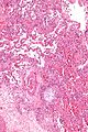
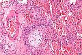
- Distal airway disease -- airways plugged with organizing exudate ("Masson bodies").
- "Organized exudate" = fluffy light-staining paucicellular regions with stellate cells (fibroblasts & immature connective tissue).
- No hobnailing of pneumocytes.
- Type 2 pneumocytes hyperplasia is absent.
DDx:
- Diffuse alveolar damage, proliferative phase - has type 2 pneumoncyte hyperplasia.
- Bronchiolitis obliterans.
Images
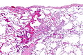
OP - very low mag. (WC/Nephron)
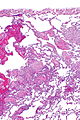
OP - low mag. (WC/Nephron)
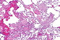
OP - low mag. (WC/Nephron)
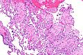
OP - intermed. mag. (WC/Nephron)
OP - intermed. mag. (WC/Nephron)
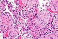
OP - high mag. (WC/Nephron)


Masson body
Masson body - intermed. mag. (WC/Nephron)
Masson body - high mag. (WC/Nephron)
Masson body - very high mag. (WC/Nephron)


www:
Sign out
Lung, Right Lower Lobe, Open Lung Biopsy:
- Organizing pneumonia.
- Emphysematous changes.
- NEGATIVE for significant pulmonary fibrosis.
- NEGATIVE for malignancy.

Comment:
There is no evidence of granulomatous disease.
Micro
Organizing pneumonia with well-formed Masson bodies is seen subpleural and peribronchial. It is pathcy and mild. Significant fibrosis is absent. Eosinophils are not readily apparent.
See also
References
- ↑ 1.0 1.1 Humphrey, Peter A; Dehner, Louis P; Pfeifer, John D (2008). The Washington Manual of Surgical Pathology (1st ed.). Lippincott Williams & Wilkins. pp. 91. ISBN 978-0781765275.
- ↑ Klatt, Edward C. (2006). Robbins and Cotran Atlas of Pathology (1st ed.). Saunders. pp. 110. ISBN 978-1416002741.