Difference between revisions of "Organizing pneumonia"
Jump to navigation
Jump to search
(→Masson body: +SO) |
|||
| (4 intermediate revisions by the same user not shown) | |||
| Line 1: | Line 1: | ||
{{ Infobox diagnosis | {{ Infobox diagnosis | ||
| Name = {{PAGENAME}} | | Name = {{PAGENAME}} | ||
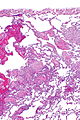
| Image = | | Image = Organizing pneumonia -- low mag.jpg | ||
| Width = | | Width = | ||
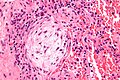
| Caption = | | Caption = Organizing pneumonia. [[H&E stain]]. | ||
| Synonyms = | | Synonyms = | ||
| Micro = | | Micro = distal airway disease -- airways plugged with organizing exudate (fluffy light-staining paucicellular regions with stellate cells); no hobnailing of pneumocytes; type 2 pneumocytes hyperplasia is absent | ||
| Subtypes = | | Subtypes = | ||
| LMDDx = | | LMDDx = [[diffuse alveolar damage]] (proliferative phase), [[bronchiolitis obliterans]]. | ||
| Stains = | | Stains = | ||
| IHC = | | IHC = | ||
| Line 61: | Line 61: | ||
===Images=== | ===Images=== | ||
<gallery> | |||
Image: Organizing pneumonia -- very low mag.jpg | OP - very low mag. (WC/Nephron) | |||
Image: Organizing pneumonia -- low mag.jpg | OP - low mag. (WC/Nephron) | |||
Image: Organizing pneumonia - alt -- low mag.jpg | OP - low mag. (WC/Nephron) | |||
Image: Organizing pneumonia - alt - intermed mag.jpg | OP - intermed. mag. (WC/Nephron) | |||
Image: Organizing pneumonia -- intermed mag.jpg | OP - intermed. mag. (WC/Nephron) | |||
Image: Organizing pneumonia -- high mag.jpg | OP - high mag. (WC/Nephron) | |||
</gallery> | |||
===Masson body=== | |||
<gallery> | <gallery> | ||
Image:Masson_body_-_intermed_mag.jpg | Masson body - intermed. mag. (WC/Nephron) | Image:Masson_body_-_intermed_mag.jpg | Masson body - intermed. mag. (WC/Nephron) | ||
| Line 67: | Line 76: | ||
</gallery> | </gallery> | ||
www: | www: | ||
*[http://casereports.bmj.com/content/2011/bcr.11.2010.3483.full BOOP (bmj.com)]. | *[http://casereports.bmj.com/content/2011/bcr.11.2010.3483.full BOOP (bmj.com)]. | ||
*[http://www.flickr.com/photos/pulmonary_pathology/4733384977/ Masson body (flickr.com)]. | *[http://www.flickr.com/photos/pulmonary_pathology/4733384977/ Masson body (flickr.com)]. | ||
*[https://www.flickr.com/photos/pulmonary_pathology/3734401023 Organizing pneumonia (flicker.com)]. | |||
==Sign out== | |||
<pre> | |||
Lung, Right Lower Lobe, Open Lung Biopsy: | |||
- Organizing pneumonia. | |||
- Emphysematous changes. | |||
- NEGATIVE for significant pulmonary fibrosis. | |||
- NEGATIVE for malignancy. | |||
Comment: | |||
There is no evidence of granulomatous disease. | |||
</pre> | |||
===Micro=== | |||
Organizing pneumonia with well-formed Masson bodies is seen subpleural and peribronchial. It is pathcy and mild. Significant fibrosis is absent. Eosinophils are not readily apparent. | |||
==See also== | ==See also== | ||
Latest revision as of 16:55, 6 April 2016
| Organizing pneumonia | |
|---|---|
| Diagnosis in short | |
 Organizing pneumonia. H&E stain. | |
|
| |
| LM | distal airway disease -- airways plugged with organizing exudate (fluffy light-staining paucicellular regions with stellate cells); no hobnailing of pneumocytes; type 2 pneumocytes hyperplasia is absent |
| LM DDx | diffuse alveolar damage (proliferative phase), bronchiolitis obliterans. |
| Site | lung - diffuse lung diseases |
|
| |
| Prevalence | uncommon |
| Prognosis | dependent on underlying cause |
| Clin. DDx | cryptogenic organizing pneumonia, transplant rejection, infection (pneumonia), collagen vascular disease, peri-tumour |
| Treatment | dependent on underlying cause |
Organizing pneumonia, abbreviated OP, is a histologic pattern in lung pathology. It fits into the larger category of diffuse lung diseases.
General
- Multiple causes, e.g. transplant rejection, infection.
Clinical diagnoses:[1]
- Transplant rejection.
- Cryptogenic organizing pneumonia (COP), AKA (idiopathic) bronchiolitis obliterans organizing pneumonia (BOOP).
- Should not be confused with constrictive bronchiolitis (AKA bronchiolitis obliterans).
- Collagen vascular disease.
- Toxic injury.
- Infection.
- Peri-tumour - in proximity to a space-occupying lesion (abscess, neoplasm).
Note:
- BOOP is used as a synonym for organizing pneumonia which has the long differential diagnosis above.[1]
- Confusingly, it may be used to refer to the idiopathic form of organizing pneumonia, now generally known as cryptogenic organizing pneumonia (COP).
- In other words, strictly speaking, BOOP is not the same as COP; idiopathic BOOP is COP.
- Confusingly, it may be used to refer to the idiopathic form of organizing pneumonia, now generally known as cryptogenic organizing pneumonia (COP).
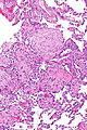
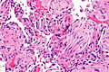
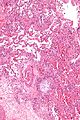
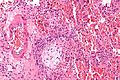
Microscopic
Features:[2]
- Distal airway disease -- airways plugged with organizing exudate ("Masson bodies").
- "Organized exudate" = fluffy light-staining paucicellular regions with stellate cells (fibroblasts & immature connective tissue).
- No hobnailing of pneumocytes.
- Type 2 pneumocytes hyperplasia is absent.
DDx:
- Diffuse alveolar damage, proliferative phase - has type 2 pneumoncyte hyperplasia.
- Bronchiolitis obliterans.
Images
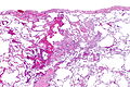
OP - very low mag. (WC/Nephron)
OP - low mag. (WC/Nephron)
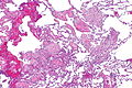
OP - low mag. (WC/Nephron)
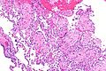
OP - intermed. mag. (WC/Nephron)
OP - intermed. mag. (WC/Nephron)
OP - high mag. (WC/Nephron)


Masson body
Masson body - intermed. mag. (WC/Nephron)
Masson body - high mag. (WC/Nephron)
Masson body - very high mag. (WC/Nephron)


www:
Sign out
Lung, Right Lower Lobe, Open Lung Biopsy:
- Organizing pneumonia.
- Emphysematous changes.
- NEGATIVE for significant pulmonary fibrosis.
- NEGATIVE for malignancy.

Comment:
There is no evidence of granulomatous disease.
Micro
Organizing pneumonia with well-formed Masson bodies is seen subpleural and peribronchial. It is pathcy and mild. Significant fibrosis is absent. Eosinophils are not readily apparent.
See also
References
- ↑ 1.0 1.1 Humphrey, Peter A; Dehner, Louis P; Pfeifer, John D (2008). The Washington Manual of Surgical Pathology (1st ed.). Lippincott Williams & Wilkins. pp. 91. ISBN 978-0781765275.
- ↑ Klatt, Edward C. (2006). Robbins and Cotran Atlas of Pathology (1st ed.). Saunders. pp. 110. ISBN 978-1416002741.