Difference between revisions of "Organizing pneumonia"
Jump to navigation
Jump to search
| Line 27: | Line 27: | ||
| Prognosis = dependent on underlying cause | | Prognosis = dependent on underlying cause | ||
| Other = | | Other = | ||
| ClinDDx = [[cryptogenic organizing pneumonia]] | | ClinDDx = [[cryptogenic organizing pneumonia]], [[lung transplant pathology|transplant rejection]], infection ([[pneumonia]]), [[collagen vascular disease]], peri-tumour | ||
| Tx = dependent on underlying cause | | Tx = dependent on underlying cause | ||
}} | }} | ||
| Line 42: | Line 42: | ||
*Toxic injury. | *Toxic injury. | ||
*Infection. | *Infection. | ||
*Peri- | *Peri-tumour - in proximity to a space-occupying lesion (abscess, neoplasm). | ||
Note: | Note: | ||
Revision as of 02:16, 31 May 2014
| Organizing pneumonia | |
|---|---|
| Diagnosis in short | |
 Masson body. H&E stain. | |
| Site | lung - diffuse lung diseases |
|
| |
| Prevalence | uncommon |
| Prognosis | dependent on underlying cause |
| Clin. DDx | cryptogenic organizing pneumonia, transplant rejection, infection (pneumonia), collagen vascular disease, peri-tumour |
| Treatment | dependent on underlying cause |
Organizing pneumonia, abbreviated OP, is a histologic pattern in lung pathology. It fits into the larger category of diffuse lung diseases.
General
- Multiple causes, e.g. transplant rejection, infection.
Clinical diagnoses:[1]
- Transplant rejection.
- Cryptogenic organizing pneumonia (COP), AKA (idiopathic) bronchiolitis obliterans organizing pneumonia (BOOP).
- Should not be confused with constrictive bronchiolitis (AKA bronchiolitis obliterans).
- Collagen vascular disease.
- Toxic injury.
- Infection.
- Peri-tumour - in proximity to a space-occupying lesion (abscess, neoplasm).
Note:
- BOOP is used as a synonym for organizing pneumonia which has the long differential diagnosis above.[1]
- Confusingly, it may be used to refer to the idiopathic form of organizing pneumonia, now generally known as cryptogenic organizing pneumonia (COP).
- In other words, strictly speaking, BOOP is not the same as COP; idiopathic BOOP is COP.
- Confusingly, it may be used to refer to the idiopathic form of organizing pneumonia, now generally known as cryptogenic organizing pneumonia (COP).
Microscopic
Features:[2]
- Distal airway disease -- airways plugged with organizing exudate ("Masson bodies").
- "Organized exudate" = fluffy light-staining paucicellular regions with stellate cells (fibroblasts & immature connective tissue).
- No hobnailing of pneumocytes.
- Type 2 pneumocytes hyperplasia is absent.
DDx:
- Diffuse alveolar damage, proliferative phase - has type 2 pneumoncyte hyperplasia.
- Bronchiolitis obliterans.
Images
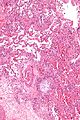
Masson body - intermed. mag. (WC/Nephron)
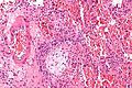
Masson body - high mag. (WC/Nephron)
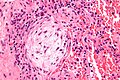
Masson body - very high mag. (WC/Nephron)


www:
See also
References
- ↑ 1.0 1.1 Humphrey, Peter A; Dehner, Louis P; Pfeifer, John D (2008). The Washington Manual of Surgical Pathology (1st ed.). Lippincott Williams & Wilkins. pp. 91. ISBN 978-0781765275.
- ↑ Klatt, Edward C. (2006). Robbins and Cotran Atlas of Pathology (1st ed.). Saunders. pp. 110. ISBN 978-1416002741.
- ↑ URL: http://150.59.224.157/pathology/index.php?first_category_id=2&second_category_id=20. Accessed on: 4 August 2011.