Oncocytoma of the salivary gland
Jump to navigation
Jump to search
The printable version is no longer supported and may have rendering errors. Please update your browser bookmarks and please use the default browser print function instead.
| Oncocytoma of the salivary gland | |
|---|---|
| Diagnosis in short | |
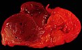
 Salivary gland oncocytoma. H&E stain. (WC/Nephron) | |
|
| |
| Synonyms | salivary gland oncocytoma |
|
| |
| LM | abundant eosinophilic cytoplasm; architecture: solid, trabecular or duct-like |
| LM DDx | acinic cell carcinoma, metastatic renal cell carcinoma, oncocytic carcinoma, granular cell tumour |
| IHC | p63 +ve |
| EM | increased numbers of mitochondria |
| Site | salivary gland |
|
| |
| Prevalence | very rare |
| Radiology | blends with normal salivary gland on T1 postcontrast and fat-saturated T2 MR images |
| Prognosis | benign |
| Clin. DDx | other salivary gland tumours |
Oncocytoma of the salivary gland (also salivary gland oncocytoma) is a rare benign tumour of the salivary glands.
General
- No risk of malignant transformation.
- Rare ~20 reported in literature.[1]
- Thought to be ~1% of all salivary gland tumours.
- Typical age: 60s-80s.
- Associated with radiation exposure.
- Major salivary glands - usually parotid gland.[2]
- Case report of oncocytoma of parotid as manifestation of Birt-Hogg-Dubé syndrome.[3]
Gross
- Golden brown appearance.
Note:
- "Disappear" into background salivary gland on MRI: isointense to normal on fat-saturated T2 and T1 postcontrast images.[4]
Image
Salivary gland oncocytoma (WC/euthman)

Microscopic
Features:
- Like oncocytomas elsewhere.
- Abundant eosinophilic cytoplasm (on H&E stain).
- Due to increased number of mitochrondria.
- Fine capillaries.
- Abundant eosinophilic cytoplasm (on H&E stain).
- Architecture: solid sheets, trabeculae or duct-like structure.[2]
Notes:
- May have clear cell change.
- Multiple small incidental lesions = oncocytosis - not oncocytoma.
DDx:
- Warthin tumour - have lymphocytes, classical bilayer.
- Acinic cell carcinoma.
- Metastatic renal cell carcinoma - esp. chromophobe renal cell carcinoma.
- Oncocytic carcinoma.
- Granular cell tumour.
Images
www:
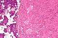
Parotid gland oncocytoma - intermed. mag. (WC/Nephron)
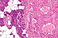
Parotid gland oncocytoma - high mag. (WC/Nephron)
Parotid gland oncocytoma - very high mag. (WC/Nephron)


IHC
EM
- Increased numbers of mitochondria.
Sign out
Nodule, Right Parotid Gland, Core Biopsy:
- ONCOCYTOMA.

Comment:
The tumour stains as follows:
POSITIVE: p63 (focal, nuclear).
NEGATIVE: CD10, RCC, S-100, CD68, vimentin.
Micro
The sections show cells with abundant eosinophilic cytoplasm and bland round nuclei in a tubular architecture. Mitotic activity is not readily apparent. Significant nuclear atypia is absent.
See also
References
- ↑ Uzunkulaoğlu H, Yazici H, Can IH, Doğan S, Uzunkulaoğlu T (May 2012). "Bilateral oncocytoma in the parotid gland". J Craniofac Surg 23 (3): e246–7. doi:10.1097/SCS.0b013e31824dfccd. PMID 22627459.
- ↑ 2.0 2.1 Zhou, CX.; Gao, Y. (Dec 2009). "Oncocytoma of the salivary glands: a clinicopathologic and immunohistochemical study.". Oral Oncol 45 (12): e232-8. doi:10.1016/j.oraloncology.2009.08.004. PMID 19796983.
- ↑ Yoshida K, Miyagawa M, Kido T, Ide K, Sano Y, Sugawara Y, Takahata H, Monden N, Furuya M, Mochizuki T (2018). "Parotid Oncocytoma as a Manifestation of Birt-Hogg-Dubé Syndrome". Case Rep Radiol 2018: 6265175. doi:10.1155/2018/6265175. PMC 6008813. PMID 29971177. https://www.ncbi.nlm.nih.gov/pmc/articles/PMC6008813/.
- ↑ Patel, ND.; van Zante, A.; Eisele, DW.; Harnsberger, HR.; Glastonbury, CM. (Oct 2011). "Oncocytoma: the vanishing parotid mass.". AJNR Am J Neuroradiol 32 (9): 1703-6. doi:10.3174/ajnr.A2569. PMID 21757520.
- ↑ 5.0 5.1 McHugh, JB.; Hoschar, AP.; Dvorakova, M.; Parwani, AV.; Barnes, EL.; Seethala, RR. (Dec 2007). "p63 immunohistochemistry differentiates salivary gland oncocytoma and oncocytic carcinoma from metastatic renal cell carcinoma.". Head Neck Pathol 1 (2): 123-31. doi:10.1007/s12105-007-0031-4. PMC 2807526. PMID 20614263. https://www.ncbi.nlm.nih.gov/pmc/articles/PMC2807526/.
- ↑ Canberk, S.; Onenerk, M.; Sayman, E.; Goret, CC.; Erkan, M.; Atasoy, T.; Kilicoglu, GZ. (2015). "Is DOG1 really useful in the diagnosis of salivary gland acinic cell carcinoma? - A DOG1 (clone K9) analysis in fine needle aspiration cell blocks and the review of the literature.". Cytojournal 12: 18. doi:10.4103/1742-6413.162774. PMID 26425134.
- ↑ Butler RT, Alderman MA, Thompson LD, McHugh JB (March 2015). "Evaluation of PAX2 and PAX8 expression in salivary gland neoplasms". Head Neck Pathol 9 (1): 47–50. doi:10.1007/s12105-014-0546-4. PMC 4382472. PMID 24771139. https://www.ncbi.nlm.nih.gov/pmc/articles/PMC4382472/.