Oncocytoma of the salivary gland
Jump to navigation
Jump to search
| Oncocytoma of the salivary gland | |
|---|---|
| Diagnosis in short | |
 Salivary gland oncocytoma. H&E stain. (WC/Nephron) | |
|
| |
| Synonyms | salivary gland oncocytoma |
|
| |
| LM | abundant eosinophilic cytoplasm; architecture: solid, trabecular or duct-like |
| LM DDx | acinic cell carcinoma, metastatic renal cell carcinoma, oncocytic carcinoma, granular cell tumour |
| IHC | p63 +ve |
| EM | increased numbers of mitochondria |
| Site | salivary gland |
|
| |
| Prevalence | very rare |
| Radiology | blends with normal salivary gland on T1 postcontrast and fat-saturated T2 MR images |
| Prognosis | benign |
| Clin. DDx | other salivary gland tumours |
Oncocytoma of the salivary gland (also salivary gland oncocytoma) is a rare benign tumour of the salivary glands.
General
- No risk of malignant transformation.
- Rare ~20 reported in literature.[1]
- Thought to be ~1% of all salivary gland tumours.
- Typical age: 60s-80s.
- Associated with radiation exposure.
- Major salivary glands - usually parotid gland.[2]
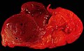
Gross
- Golden brown appearance.
Note:
- "Disappear" into background salivary gland on MRI: isointense to normal on fat-saturated T2 and T1 postcontrast images.[3]
Image
Salivary gland oncocytoma (WC/euthman)

Microscopic
Features:
- Like oncocytomas elsewhere.
- Abundant eosinophilic cytoplasm (on H&E stain).
- Due to increased number of mitochrondria.
- Fine capillaries.
- Abundant eosinophilic cytoplasm (on H&E stain).
- Architecture: solid sheets, trabeculae or duct-like structure.[2]
Notes:
- May have clear cell change.
- Multiple small incidental lesions = oncocytosis - not oncocytoma.
DDx:
- Acinic cell carcinoma.
- Metastatic renal cell carcinoma - esp. chromophobe renal cell carcinoma.
- Oncocytic carcinoma.
- Granular cell tumour.
Images
www:
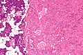
Parotid gland oncocytoma - intermed. mag. (WC/Nephron)
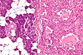
Parotid gland oncocytoma - high mag. (WC/Nephron)
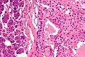
Parotid gland oncocytoma - very high mag. (WC/Nephron)


IHC
EM
- Increased numbers of mitochondria.
Sign out
Nodule, Right Parotid Gland, Core Biopsy:
- ONCOCYTOMA.

Comment:
The tumour stains as follows:
POSITIVE: p63 (focal, nuclear).
NEGATIVE: CD10, RCC, S-100, CD68, vimentin.
Micro
The sections show cells with abundant eosinophilic cytoplasm and bland round nuclei in a tubular architecture. Mitotic activity is not readily apparent. Significant nuclear atypia is absent.
See also
References
- ↑ Uzunkulaoğlu H, Yazici H, Can IH, Doğan S, Uzunkulaoğlu T (May 2012). "Bilateral oncocytoma in the parotid gland". J Craniofac Surg 23 (3): e246–7. doi:10.1097/SCS.0b013e31824dfccd. PMID 22627459.
- ↑ 2.0 2.1 Zhou, CX.; Gao, Y. (Dec 2009). "Oncocytoma of the salivary glands: a clinicopathologic and immunohistochemical study.". Oral Oncol 45 (12): e232-8. doi:10.1016/j.oraloncology.2009.08.004. PMID 19796983.
- ↑ Patel, ND.; van Zante, A.; Eisele, DW.; Harnsberger, HR.; Glastonbury, CM. (Oct 2011). "Oncocytoma: the vanishing parotid mass.". AJNR Am J Neuroradiol 32 (9): 1703-6. doi:10.3174/ajnr.A2569. PMID 21757520.
- ↑ 4.0 4.1 McHugh, JB.; Hoschar, AP.; Dvorakova, M.; Parwani, AV.; Barnes, EL.; Seethala, RR. (Dec 2007). "p63 immunohistochemistry differentiates salivary gland oncocytoma and oncocytic carcinoma from metastatic renal cell carcinoma.". Head Neck Pathol 1 (2): 123-31. doi:10.1007/s12105-007-0031-4. PMC 2807526. PMID 20614263. https://www.ncbi.nlm.nih.gov/pmc/articles/PMC2807526/.
- ↑ Canberk, S.; Onenerk, M.; Sayman, E.; Goret, CC.; Erkan, M.; Atasoy, T.; Kilicoglu, GZ. (2015). "Is DOG1 really useful in the diagnosis of salivary gland acinic cell carcinoma? - A DOG1 (clone K9) analysis in fine needle aspiration cell blocks and the review of the literature.". Cytojournal 12: 18. doi:10.4103/1742-6413.162774. PMID 26425134.