Nucleolus
Jump to navigation
Jump to search
The printable version is no longer supported and may have rendering errors. Please update your browser bookmarks and please use the default browser print function instead.

The nucleolus (plural nucleoli) is a thingy in the nucleus that may give the pathologist a clue to what they are looking at.
Generally speaking, large nucleoli suggest something is happening - they are associated with gene transcription. Large nucleoli are seen in malignancies and reactive conditions.
Macronucleolus
Almost the size of RBC ~ 6-7 micrometers.
Example:
- Reed-Sternberg cell (Hodgkin lymphoma) ~ 5-7 micrometers.[1]
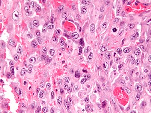
Image
Extreme nuclear enlargement with huge macronucleoli. (WC)
Red nucleolus
Large - can be seen with 10x objective.
Examples:
- Melanoma.
- Serous carcinoma.
- Hereditary leiomyomatosis renal cell carcinoma syndrome associated renal cell carcinoma.
Large nucleolus
Large - can be seen with 10x objective.
Examples:
- Melanoma.
- Carcinoma.
- Serous carcinoma.
- Adenocarcinoma.
- High-grade renal cell carcinoma.
- Sarcoma:
- Ganglion cell:
- Ganglion (benign).
- Gangliocytic paraganglioma.
- Ganglioneuroma.
Medium-sized nucleolus
Medium - can be seen well with 20x objective.
Examples:
- Prostatic adenocarcinoma.
- Oncocytoma.
- Mammary carcinoma, no special type.
- Embryonal carcinoma.
- Squamous metaplasia of the uterine cervix.
Small
Small - hard to see at 20x objective, seen with 40x objective.
Examples:
Indistinct nucleolus
Not present - cannot see with 40x objective.
Examples:
See also
References
- ↑ Mitchell, Richard; Kumar, Vinay; Fausto, Nelson; Abbas, Abul K.; Aster, Jon (2011). Pocket Companion to Robbins & Cotran Pathologic Basis of Disease (8th ed.). Elsevier Saunders. pp. 329. ISBN 978-1416054542.