Neurocytoma
Jump to navigation
Jump to search
Neurocytoma is a rare neuropathology tumour.
General
- Rare.
Microscopic
Features:[1]
- Pineocytomatous/neurocytic rosette = irregular rosette with a large meshwork of fibers (neuropil) at the centre.[2]
- Similar to Homer-Wright rosette.
- Monomorphic cells with round nuclei and speckled chromatin.
- Perinuclear clearing.
- Ganglion cell differentiation.
- Well-defined cell borders.
- Hyalinized vessels.
- Necrosis absent.
IHC
- MIB-1: Usu. low.
- Synapthophysin +ve.
DDx:
- Oligodendroglioma - do not have the characteristic rosettes.
- Ganglioglioma.
- Ependymoma.
- Pilocytic astrocytoma (with predominantly oligo-like cell component).
- Diffuse leptomeningeal gliomeuronal tumour.
Images
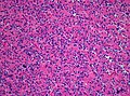
Neurocytoma (H&E: WC, Marvin101).
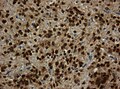
Nuclear NeuN immunoreactivity (WC, Marvin101):


www:
IHC
- Synaptophysin +ve.
- Most glial tumour -ve.[3]
Molecular
- FGFR1-TACC1 fusions common.[4]
See also
References
- ↑ URL: http://moon.ouhsc.edu/kfung/jty1/Composites/FNA0IE14-Neurocytoma-Micro.htm. Accessed on: 12 October 2011.
- ↑ Wippold FJ, Perry A (March 2006). "Neuropathology for the neuroradiologist: rosettes and pseudorosettes". AJNR Am J Neuroradiol 27 (3): 488–92. PMID 16551982.
- ↑ URL: http://path.upmc.edu/cases/case383/dx.html. Accessed on: 15 January 2012.
- ↑ Sievers, P.; Stichel, D.; Schrimpf, D.; Sahm, F.; Koelsche, C.; Reuss, DE.; Wefers, AK.; Reinhardt, A. et al. (Jul 2018). "FGFR1:TACC1 fusion is a frequent event in molecularly defined extraventricular neurocytoma.". Acta Neuropathol. doi:10.1007/s00401-018-1882-3. PMID 29978331.