Difference between revisions of "Microcystic elongated and fragmented glands in endometrioid endometrial carcinoma"
Jump to navigation
Jump to search

(+image) |
(→Microscopic: more images) |
||
| Line 12: | Line 12: | ||
*[[Lymphovascular invasion]] more commmon than in non-MELF carcinomas.<ref name=pmid24487466>{{cite journal |vauthor=Hertel JD, Huettner PC, Pfeifer JD |title=Lymphovascular space invasion in microcystic elongated and fragmented (MELF)-pattern well-differentiated endometrioid adenocarcinoma is associated with a higher rate of lymph node metastasis |journal=Int. J. Gynecol. Pathol. |volume=33 |issue=2 |pages=127–34 |date=March 2014 |pmid=24487466 |doi=10.1097/PGP.0b013e318285657b |url=}}</ref> | *[[Lymphovascular invasion]] more commmon than in non-MELF carcinomas.<ref name=pmid24487466>{{cite journal |vauthor=Hertel JD, Huettner PC, Pfeifer JD |title=Lymphovascular space invasion in microcystic elongated and fragmented (MELF)-pattern well-differentiated endometrioid adenocarcinoma is associated with a higher rate of lymph node metastasis |journal=Int. J. Gynecol. Pathol. |volume=33 |issue=2 |pages=127–34 |date=March 2014 |pmid=24487466 |doi=10.1097/PGP.0b013e318285657b |url=}}</ref> | ||
*Mucinous differentiation common in MELF.<ref name=pmid19900084/> | *Mucinous differentiation common in MELF.<ref name=pmid19900084/> | ||
===Images=== | |||
<gallery> | |||
Image:Endometrioid endometrial adenocarcinoma with MELF -- very low mag.jpg | EEAd w/ MELF - very low mag. (WC) | |||
Image:Endometrioid endometrial adenocarcinoma with MELF - alt -- low mag.jpg | EEAd w/ MELF - low mag. (WC) | |||
Image:Endometrioid endometrial adenocarcinoma with MELF -- intermed mag.jpg | EEAd w/ MELF - intermed. mag. (WC) | |||
Image:Endometrioid endometrial adenocarcinoma with MELF - alt -- intermed mag.jpg | EEAd w/ MELF - intermed. mag. (WC) | |||
Image:Endometrioid endometrial adenocarcinoma with MELF - alt2 -- intermed mag.jpg | EEAd w/ MELF - intermed. mag. (WC) | |||
Image:Endometrioid endometrial adenocarcinoma with MELF -- high mag.jpg | EEAd w/ MELF - high mag. (WC) | |||
</gallery> | |||
==See also== | ==See also== | ||
Latest revision as of 04:59, 24 May 2020

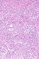
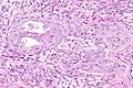
Micrograph showing endometrioid endometrial adenocarcinoma with microcystic elongated and fragmented (MELF) glands. H&E stain.
Microcystic elongated and fragmented glands in endometrioid endometrial carcinoma is a pattern of endometrioid endometrial carcinoma. Microcystic elongated and fragmented glands is typically abbreviated as MELF.[1]
General
- MELF pattern is a poor prognosticator.[2]
Microscopic
Features:
- Irregular elongated glands ("microcystic elongated and fragmented") - at deep aspect of tumour.
- Surrounded by (pale) myxoid stroma.[3]
Notes:
- Lymphovascular invasion more commmon than in non-MELF carcinomas.[2]
- Mucinous differentiation common in MELF.[3]
Images
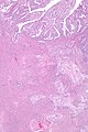
EEAd w/ MELF - very low mag. (WC)
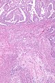
EEAd w/ MELF - low mag. (WC)
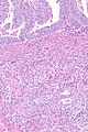
EEAd w/ MELF - intermed. mag. (WC)
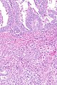
EEAd w/ MELF - intermed. mag. (WC)
EEAd w/ MELF - intermed. mag. (WC)
EEAd w/ MELF - high mag. (WC)


See also
References
- ↑ Euscher, E.; Fox, P.; Bassett, R.; Al-Ghawi, H.; Ali-Fehmi, R.; Barbuto, D.; Djordjevic, B.; Frauenhoffer, E. et al. (Nov 2013). "The pattern of myometrial invasion as a predictor of lymph node metastasis or extrauterine disease in low-grade endometrial carcinoma.". Am J Surg Pathol 37 (11): 1728-36. doi:10.1097/PAS.0b013e318299f2ab. PMID 24061515.
- ↑ 2.0 2.1 "Lymphovascular space invasion in microcystic elongated and fragmented (MELF)-pattern well-differentiated endometrioid adenocarcinoma is associated with a higher rate of lymph node metastasis". Int. J. Gynecol. Pathol. 33 (2): 127–34. March 2014. doi:10.1097/PGP.0b013e318285657b. PMID 24487466.
- ↑ 3.0 3.1 Stewart CJ, Brennan BA, Leung YC, Little L (2009). "MELF pattern invasion in endometrial carcinoma: association with low grade, myoinvasive endometrioid tumours, focal mucinous differentiation and vascular invasion". Pathology 41 (5): 454–9. doi:10.1080/00313020903041135. PMID 19900084.