Mesenchymal chondrosarcoma
Jump to navigation
Jump to search
The printable version is no longer supported and may have rendering errors. Please update your browser bookmarks and please use the default browser print function instead.
Mesenchymal chondrosarcoma is a rare type of chondrosarcoma that is found in the soft tissue.
General
- Rare variant of chondrosarcoma.
- 2–10% of primary chondrosarcomas.
- Adolescents and young adults.
- Female predilection.
- Most commonly intraosseous but can occur in extraskeletal sites especially the central nervous system (from the meninges).
- The mesenchymal in the name refers to the ability to arise in soft tissues.[1]
- Conceptualized as originating from a pleuripotential mesenchymal cell with foci recapitulating enchondral ossification.
- The small cells appear to be an undifferentiated cartilage stem cell which “differentiate” into benign cartilage.[2]
Gross
Pink and fleshy with foci of calcification.
Microscopic
Features:
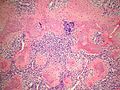
- Malignant tumour with a characteristic biphasic pattern.
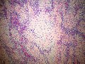
- Cellular poorly differentiated small round blue cells.
- Islands of well-differentiated hyaline cartilage.
- Progressive maturation of cartilage towards the center.
- Central calcification or bone formation.
- Can have a hemangiopericytomatous vascular pattern.
Notes:
- May be described as white clouds in a dark blue sky.
DDX
- Hemangiopericytoma - no cartilage.
- Lymphoma - Sox9 negative, CD45 positive.
- Metaplastic glioblastoma - usually older adults - GFAP positive.
- Chondrosarcoma (NOS) - usually older adults - hyaline cartilage is malignant.
- Small cell osteosarcoma - Sox10 negative, no cartilage.
- Ewing sarcoma - both are CD99 positive but ES is Sox9 negative, no cartilage.
- Monophasic synovial sarcoma - also can have the hemangiopericytomatous vasculature.
Note:
- Depends a bit on where the tumour is located and how much cartilage is readily visible.
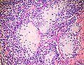
Images
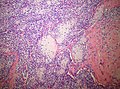
Islands of cartilage in a background of small blue cells.(SKB)
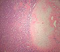
Islands of cartilage in a background of small blue cells.(SKB)
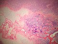
Islands of cartilage in a background of small blue cells.(SKB)
Islands of cartilage in a background of small blue cells.(SKB)
Islands of cartilage in a background of small blue cells.(SKB)
Islands of cartilage in a background of small blue cells.(SKB)

Small blue cells predominate.(SKB)


www:
- Mesenchymal chondrosarcoma (ouhsc.edu).
- Pathology Outlines - Hemangiopericytomatous example [1]
- Sarcoma Images - [2]
- CNS Atlas - [3]
- CNS Atlas - [4]
- CNS Atlas - [5]
![[1]](http://pathologyoutlines.com/wick/chondrosarcoma%20mesenchymal%20type%20micro0007.jpg)
![[3]](http://www.cnsatlas.com/medicalatlas/images/370/800_poIB6FYZQ73wh.jpg)
![[4]](http://www.cnsatlas.com/medicalatlas/images/370/800_9oJtTQXXxaEB3.jpg)
![[5]](http://www.cnsatlas.com/medicalatlas/images/370/800_8h2C7upeOj3EB.jpg)
IHC
- SOX9 (positive in small cells and chondrocytes).[3]
- S100 (positive in chondrocytes not in small cells).
- Osteocalcin (negative in small cells).
- CD99 - (positive in small cells)
Molecular
See also
References
- ↑ Dowling EA (June 1964). "Mesenchymal chondrosarcoma". J Bone Joint Surg Am 46: 747–54. PMID 14161087. http://www.ejbjs.org/cgi/reprint/46/4/747.pdf.
- ↑ Fanburg-Smith, JC.; Auerbach, A.; Marwaha, JS.; Wang, Z.; Rushing, EJ. (May 2010). "Reappraisal of mesenchymal chondrosarcoma: novel morphologic observations of the hyaline cartilage and endochondral ossification and beta-catenin, Sox9, and osteocalcin immunostaining of 22 cases.". Hum Pathol 41 (5): 653-62. doi:10.1016/j.humpath.2009.11.006. PMID 20138330.
- ↑ Pang, ZG.; He, XZ.; Wu, LY.; Wei, W.; Liu, XY.; Liao, DY.; Li, FY.; Zhang, XL. (Jun 2011). "[Clinicopathologic and immunohistochemical study of 23 cases of mesenchymal chondrosarcoma].". Zhonghua Bing Li Xue Za Zhi 40 (6): 368-72. PMID 21914343.
- ↑ Panagopoulos, I.; Gorunova, L.; Bjerkehagen, B.; Boye, K.; Heim, S. (Jul 2014). "Chromosome aberrations and HEY1-NCOA2 fusion gene in a mesenchymal chondrosarcoma.". Oncol Rep 32 (1): 40-4. doi:10.3892/or.2014.3180. PMID 24839999.
- ↑ Nyquist KB, Panagopoulos I, Thorsen J, Haugom L, Gorunova L, Bjerkehagen B, Fosså A, Guriby M, Nome T, Lothe RA, Skotheim RI, Heim S, Micci F (2012). "Whole-transcriptome sequencing identifies novel IRF2BP2-CDX1 fusion gene brought about by translocation t(1;5)(q42;q32) in mesenchymal chondrosarcoma". PLoS ONE 7 (11): e49705. doi:10.1371/journal.pone.0049705. PMC 3504151. PMID 23185413. https://www.ncbi.nlm.nih.gov/pmc/articles/PMC3504151/.