Difference between revisions of "Malignant mesothelioma"
Jump to navigation
Jump to search
(create +cat.) |
(split out) |
||
| Line 1: | Line 1: | ||
{{ Infobox diagnosis | |||
| Name = {{PAGENAME}} | |||
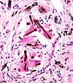
| Image = Malignant epithelioid mesothelioma - high mag.jpg | |||
| Width = | |||
| Caption = Malignant mesothelioma. [[H&E stain]]. | |||
| Micro = infiltrative atypical cells (epithelioid, spindled or both) | |||
| Subtypes = biphasic mesothelioma, epithelioid mesothelioma, desmoplastic mesothelioma, sarcomatoid mesothelioma. | |||
| LMDDx = mesothelial hyperplasia, fibrosing pleuritis, [[adenocarcinoma]] - esp. [[lung adenocarcinoma|lung]] | |||
| Stains = | |||
| IHC = calretinin +ve, D2-40 +ve, CK5/6 +ve, WT-1 +ve, CK7 +ve, CEA -ve, TTF-1 -ve | |||
| EM = | |||
| Molecular = | |||
| IF = | |||
| Gross = | |||
| Grossing = | |||
| Site = [[lung]], peritoneum | |||
| Assdx = | |||
| Syndromes = | |||
| Clinicalhx = asbestos exposure, smoking | |||
| Signs = | |||
| Symptoms = | |||
| Prevalence = rare | |||
| Bloodwork = | |||
| Rads = | |||
| Endoscopy = | |||
| Prognosis = very poor | |||
| Other = | |||
| ClinDDx = | |||
}} | |||
'''Malignant mesothelioma''', also '''mesothelioma''', is a form of [[cancer]] that arise from the mesothelial lining. | |||
It should '''not''' be confused with ''[[benign multicystic mesothelioma]]'' and ''[[benign papillary mesothelioma]]''. | |||
==General== | |||
*Poor prognosis - median survival <12 months.<ref name=pmid23413596>{{Cite journal | last1 = Mineo | first1 = TC. | last2 = Ambrogi | first2 = V. | title = Malignant pleural mesothelioma: factors influencing the prognosis. | journal = Oncology (Williston Park) | volume = 26 | issue = 12 | pages = 1164-75 | month = Dec | year = 2012 | doi = | PMID = 23413596 }}</ref> | |||
Locations: | |||
*Lung. | |||
*Primary peritoneal. | |||
Epidemiology: | |||
*Strong association with asbestos exposure. | |||
Conditions associated with asbestos exposure (mnemonic ''PALM''):<ref name=Ref_PCPBoD8_375>{{Ref PCPBoD8|375}}</ref> | |||
*Pleural plaques. | |||
*[[Asbestosis]]. | |||
*[[Lung carcinoma]]. | |||
*Malignant mesothelioma. | |||
Possible association with asbestos exposure: | |||
*[[Gestational trophoblastic disease]].<ref name=pmid19900938>{{Cite journal | last1 = Reid | first1 = A. | last2 = Heyworth | first2 = J. | last3 = de Klerk | first3 = N. | last4 = Musk | first4 = AW. | title = Asbestos exposure and gestational trophoblastic disease: a hypothesis. | journal = Cancer Epidemiol Biomarkers Prev | volume = 18 | issue = 11 | pages = 2895-8 | month = Nov | year = 2009 | doi = 10.1158/1055-9965.EPI-09-0731 | PMID = 19900938 }}</ref> | |||
==Microscopic== | |||
Features:<ref name=Ref_WMSP156>{{Ref WMSP|156}}</ref> | |||
*Infiltrative atypical cells - '''key feature'''. | |||
**+/-Epithelioid cells - may be cytologically bland, i.e. benign appearing. | |||
***Variable architecture: sheets, microglandular, tubulopapillary. | |||
***+/-[[Psammoma bodies]]. | |||
**+/-Spindle cells. | |||
*+/-''Ferruginous body'' - '''strongly supportive'''.<ref>URL: [http://medical-dictionary.thefreedictionary.com/asbestos+body http://medical-dictionary.thefreedictionary.com/asbestos+body]. Accessed on: 4 November 2011.</ref> | |||
** Looks like a (twirling) baton - segemented appearance, brown colour. | |||
** Thin (asbestos) fiber in the core. | |||
Note: | |||
*''Asbestos body'' is not strictly speaking a synonym for ''ferruginous body''. | |||
DDx:<ref name=pmid15559051>{{Cite journal | last1 = Corson | first1 = JM. | title = Pathology of mesothelioma. | journal = Thorac Surg Clin | volume = 14 | issue = 4 | pages = 447-60 | month = Nov | year = 2004 | doi = 10.1016/j.thorsurg.2004.06.007 | PMID = 15559051 }} | |||
</ref> | |||
*[[Fibrosing pleuritis]]. | |||
*Mesothelial hyperplasia. | |||
===Image=== | |||
<gallery> | |||
Image:Ferruginous_body.jpg | Ferruginous body. (WC) | |||
</gallery> | |||
===Subtypes=== | |||
List of subtypes - mnemonic ''BEDS'':<ref name=pmid15559051/><ref name=Ref_WMSP156>{{Ref WMSP|156}}</ref> | |||
*Biphasic mesothelioma. | |||
**10%+ of epithelioid & 10%+ sarcomatoid. | |||
*Epithelioid mesothelioma. | |||
*Desmoplastic mesothelioma. | |||
**Should be 50%+ dense tissue with storiform pattern & atypical cells. | |||
*Sarcomatoid mesothelioma. | |||
==Stains== | |||
*PASD -ve. | |||
*Mucicarmine -ve. | |||
**Typically +ve in adenocarcinoma. | |||
==IHC== | |||
===Mesothelioma versus mesothelial hyperplasia=== | |||
Features:<ref name=pmid20209622>{{Cite journal | last1 = Hasteh | first1 = F. | last2 = Lin | first2 = GY. | last3 = Weidner | first3 = N. | last4 = Michael | first4 = CW. | title = The use of immunohistochemistry to distinguish reactive mesothelial cells from malignant mesothelioma in cytologic effusions. | journal = Cancer Cytopathol | volume = 118 | issue = 2 | pages = 90-6 | month = Apr | year = 2010 | doi = 10.1002/cncy.20071 | PMID = 20209622 }}</ref> | |||
*EMA +ve ~100% (vs. ~10%). | |||
*Desmin -ve ~5% (vs. ~85%). | |||
*GLUT1 +ve ~50% (vs. ~10%) | |||
*p53 +ve ~50% (vs. ~2%). | |||
===Mesothelioma versus adenocarcinoma=== | |||
*Several panel exists - ''no agreed upon best panel''.<ref name=pmid18318582>{{cite journal |author=Marchevsky AM |title=Application of immunohistochemistry to the diagnosis of malignant mesothelioma |journal=Arch. Pathol. Lab. Med. |volume=132 |issue=3 |pages=397-401 |year=2008 |month=March |pmid=18318582 |doi= |url=http://journals.allenpress.com/jrnlserv/?request=get-abstract&issn=0003-9985&volume=132&page=397}}</ref> | |||
**Usually two carcinoma markers + two mesothelial markers. | |||
Panel:<ref name=pmid18318582/> | |||
*Mesothelial markers: | |||
**Calretinin. | |||
**WT-1. | |||
**D2-40. | |||
**CK5/6. | |||
*Carcinoma markers: | |||
**CEA (monoclonal and polyclonal). | |||
**TTF-1. | |||
**Ber-EP4. | |||
**MOC-31. | |||
**CD15. | |||
==See also== | |||
*[[Lung tumours]]. | |||
==References== | |||
{{Reflist|2}} | |||
[[Category:Diagnosis]] | [[Category:Diagnosis]] | ||
Revision as of 22:51, 11 July 2013
| Malignant mesothelioma | |
|---|---|
| Diagnosis in short | |
 Malignant mesothelioma. H&E stain. | |
|
| |
| LM | infiltrative atypical cells (epithelioid, spindled or both) |
| Subtypes | biphasic mesothelioma, epithelioid mesothelioma, desmoplastic mesothelioma, sarcomatoid mesothelioma. |
| LM DDx | mesothelial hyperplasia, fibrosing pleuritis, adenocarcinoma - esp. lung |
| IHC | calretinin +ve, D2-40 +ve, CK5/6 +ve, WT-1 +ve, CK7 +ve, CEA -ve, TTF-1 -ve |
| Site | lung, peritoneum |
|
| |
| Clinical history | asbestos exposure, smoking |
| Prevalence | rare |
| Prognosis | very poor |
Malignant mesothelioma, also mesothelioma, is a form of cancer that arise from the mesothelial lining.
It should not be confused with benign multicystic mesothelioma and benign papillary mesothelioma.
General
- Poor prognosis - median survival <12 months.[1]
Locations:
- Lung.
- Primary peritoneal.
Epidemiology:
- Strong association with asbestos exposure.
Conditions associated with asbestos exposure (mnemonic PALM):[2]
- Pleural plaques.
- Asbestosis.
- Lung carcinoma.
- Malignant mesothelioma.
Possible association with asbestos exposure:
Microscopic
Features:[4]
- Infiltrative atypical cells - key feature.
- +/-Epithelioid cells - may be cytologically bland, i.e. benign appearing.
- Variable architecture: sheets, microglandular, tubulopapillary.
- +/-Psammoma bodies.
- +/-Spindle cells.
- +/-Epithelioid cells - may be cytologically bland, i.e. benign appearing.
- +/-Ferruginous body - strongly supportive.[5]
- Looks like a (twirling) baton - segemented appearance, brown colour.
- Thin (asbestos) fiber in the core.
Note:
- Asbestos body is not strictly speaking a synonym for ferruginous body.
DDx:[6]
- Fibrosing pleuritis.
- Mesothelial hyperplasia.
Image
Ferruginous body. (WC)

Subtypes
List of subtypes - mnemonic BEDS:[6][4]
- Biphasic mesothelioma.
- 10%+ of epithelioid & 10%+ sarcomatoid.
- Epithelioid mesothelioma.
- Desmoplastic mesothelioma.
- Should be 50%+ dense tissue with storiform pattern & atypical cells.
- Sarcomatoid mesothelioma.
Stains
- PASD -ve.
- Mucicarmine -ve.
- Typically +ve in adenocarcinoma.
IHC
Mesothelioma versus mesothelial hyperplasia
Features:[7]
- EMA +ve ~100% (vs. ~10%).
- Desmin -ve ~5% (vs. ~85%).
- GLUT1 +ve ~50% (vs. ~10%)
- p53 +ve ~50% (vs. ~2%).
Mesothelioma versus adenocarcinoma
- Several panel exists - no agreed upon best panel.[8]
- Usually two carcinoma markers + two mesothelial markers.
Panel:[8]
- Mesothelial markers:
- Calretinin.
- WT-1.
- D2-40.
- CK5/6.
- Carcinoma markers:
- CEA (monoclonal and polyclonal).
- TTF-1.
- Ber-EP4.
- MOC-31.
- CD15.
See also
References
- ↑ Mineo, TC.; Ambrogi, V. (Dec 2012). "Malignant pleural mesothelioma: factors influencing the prognosis.". Oncology (Williston Park) 26 (12): 1164-75. PMID 23413596.
- ↑ Mitchell, Richard; Kumar, Vinay; Fausto, Nelson; Abbas, Abul K.; Aster, Jon (2011). Pocket Companion to Robbins & Cotran Pathologic Basis of Disease (8th ed.). Elsevier Saunders. pp. 375. ISBN 978-1416054542.
- ↑ Reid, A.; Heyworth, J.; de Klerk, N.; Musk, AW. (Nov 2009). "Asbestos exposure and gestational trophoblastic disease: a hypothesis.". Cancer Epidemiol Biomarkers Prev 18 (11): 2895-8. doi:10.1158/1055-9965.EPI-09-0731. PMID 19900938.
- ↑ 4.0 4.1 Humphrey, Peter A; Dehner, Louis P; Pfeifer, John D (2008). The Washington Manual of Surgical Pathology (1st ed.). Lippincott Williams & Wilkins. pp. 156. ISBN 978-0781765275.
- ↑ URL: http://medical-dictionary.thefreedictionary.com/asbestos+body. Accessed on: 4 November 2011.
- ↑ 6.0 6.1 Corson, JM. (Nov 2004). "Pathology of mesothelioma.". Thorac Surg Clin 14 (4): 447-60. doi:10.1016/j.thorsurg.2004.06.007. PMID 15559051.
- ↑ Hasteh, F.; Lin, GY.; Weidner, N.; Michael, CW. (Apr 2010). "The use of immunohistochemistry to distinguish reactive mesothelial cells from malignant mesothelioma in cytologic effusions.". Cancer Cytopathol 118 (2): 90-6. doi:10.1002/cncy.20071. PMID 20209622.
- ↑ 8.0 8.1 Marchevsky AM (March 2008). "Application of immunohistochemistry to the diagnosis of malignant mesothelioma". Arch. Pathol. Lab. Med. 132 (3): 397-401. PMID 18318582. http://journals.allenpress.com/jrnlserv/?request=get-abstract&issn=0003-9985&volume=132&page=397.