Lung metastasis
Jump to navigation
Jump to search
The printable version is no longer supported and may have rendering errors. Please update your browser bookmarks and please use the default browser print function instead.
| Lung metastasis | |
|---|---|
| Diagnosis in short | |
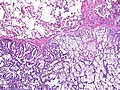
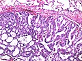
 Lung metastasis (Ewing sarcoma). H&E stain. | |
| LM DDx | primary lung cancer (adenocarcinoma of the lung, squamous cell carcinoma of the lung, small cell carcinoma of the lung), pulmonary meningothelial-like nodule, carcinoid tumourlet, carcinoid lung tumour |
| IHC | TTF-1 (-ve useful if non-squamous), CK20 (+ve suggestive colorectal carcinoma), CK7 (-ve useful if non-squamous), GATA3 (+ve suggestive UCC) |
| Gross | lung nodules - typically multiple and peripheral |
| Site | lung |
|
| |
| Clinical history | +/-history of malignancy |
| Prevalence | relatively common |
| Radiology | peripheral lung lesions, typically multiple |
| Prognosis | usually poor |
| Clin. DDx | lung primary, abscess, multiple benign lung tumours as may be seen in DIPNECH |
| Treatment | dependent on primary site, occasionally surgical |
Lung metastasis, also pulmonary metastasis and metastatic lung disease, is relatively common and generally carries a poor prognosis.
General
- Relatively common.
Gross
- Typically peripheral, multiple, well-circumscribed & white/tan masses.
- May be diffuse without an obvious mass +/- septal thickening.
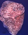
Prostate carcinoma. (WC/Rosen)
.jpg)
Microscopic
Features:
- Variable - dependent on site of origin.
- Colorectal adenocarcinoma - usually distinctive morphologically:
- Typically gland forming.
- Ellipsoid/elongated pseudostratified nuclei with moderate nuclear atypia.
- +/-Dirty necrosis.
- Typically gland forming.
- Others:
- Urothelial carcinoma - may mimic squamous cell carcinoma of the lung.
- Upper GI adenocarcinoma (e.g. gastric adenocarcinoma) - may mimic lung adenocarcinoma.
- Breast carcinoma - esp. ductal carcinoma of the breast - may mimic lung adenocarcinoma.
DDx:
- Primary lung tumour, e.g. lung adenocarcinoma, lung squamous cell carcinoma, small cell carcinoma of the lung.
- Pulmonary meningothelial-like nodule.[1]
- Carcinoid tumourlet.
- Carcinoid lung tumour.
Images
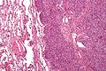
Lung metastasis (ES) - intermed. mag. (WC/Nephron)
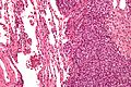
Lung metastasis (ES) - high mag. (WC/Nephron)
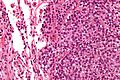
Lung metastasis (ES) - very high mag. (WC/Nephron)


Prostate carcinoma. (WC/Rosen)
Prostate carcinoma. (WC/Rosen)
.jpg)
.jpg)
IHC
- TTF-1 -ve/+ve.
- Negative suggestive of metastasis... unless it is squamous carcinoma.
- CK20 +ve/-ve.
- Positive in colorectal carcinoma - very useful.
- Negative in lung primaries.
- GATA3 +ve/-ve.
- Usu. +ve in urothelial carcinoma.
- Negative in lung primaries.[2]
- CK7 -ve/+ve.
- Positive in lung adenocarcinoma and small carcinoma of the lung.
- Positive in a number of other tumours - breast, upper GI tract, thyroid, mesothelioma, salivary gland.
- Negative in poorly differentiated carcinoma of the lung and squamous carcinoma of the lung.
See also
References
- ↑ Kfoury, H.; Arafah, MA.; Arafah, MM.; Alnassar, S.; Hajjar, W. (Feb 2012). "Mimicry of Minute Pulmonary Meningothelial-like Nodules to Metastatic Deposits in a Patient with Infiltrating Lobular Carcinoma: A Case Report and Review of the Literature.". Korean J Pathol 46 (1): 87-91. doi:10.4132/KoreanJPathol.2012.46.1.87. PMID 23109985.
- ↑ Chang, A.; Amin, A.; Gabrielson, E.; Illei, P.; Roden, RB.; Sharma, R.; Epstein, JI. (Oct 2012). "Utility of GATA3 immunohistochemistry in differentiating urothelial carcinoma from prostate adenocarcinoma and squamous cell carcinomas of the uterine cervix, anus, and lung.". Am J Surg Pathol 36 (10): 1472-6. doi:10.1097/PAS.0b013e318260cde7. PMID 22982890.