Difference between revisions of "Lung bleb"
Jump to navigation
Jump to search
| Line 1: | Line 1: | ||
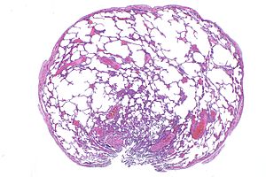
[[Image:Lung bleb -- extremely low mag.jpg|thumb|right|Lung bleb. [[H&E stain]].]] | |||
'''Lung bleb''', also '''pulmonary bleb''', are benign cystic [[lung]] lesions.<ref name=pmid12934786>{{Cite journal | last1 = Ryu | first1 = JH. | last2 = Swensen | first2 = SJ. | title = Cystic and cavitary lung diseases: focal and diffuse. | journal = Mayo Clin Proc | volume = 78 | issue = 6 | pages = 744-52 | month = Jun | year = 2003 | doi = 10.4065/78.6.744 | PMID = 12934786 }}</ref> | '''Lung bleb''', also '''pulmonary bleb''', are benign cystic [[lung]] lesions.<ref name=pmid12934786>{{Cite journal | last1 = Ryu | first1 = JH. | last2 = Swensen | first2 = SJ. | title = Cystic and cavitary lung diseases: focal and diffuse. | journal = Mayo Clin Proc | volume = 78 | issue = 6 | pages = 744-52 | month = Jun | year = 2003 | doi = 10.4065/78.6.744 | PMID = 12934786 }}</ref> | ||
| Line 23: | Line 24: | ||
*[[Emphysema]]. | *[[Emphysema]]. | ||
*[[Lymphangioleiomyomatosis]] (LAM). | *[[Lymphangioleiomyomatosis]] (LAM). | ||
===Images=== | |||
<gallery> | |||
Image:Lung bleb -- extremely low mag.jpg | Lung bleb - extremely low mag. (WC) | |||
</gallery> | |||
==Sign out== | ==Sign out== | ||
Revision as of 03:47, 3 January 2017

Lung bleb. H&E stain.
Lung bleb, also pulmonary bleb, are benign cystic lung lesions.[1]
Lung bulla and lung bullae redirect to this article.
General
- Benign.
- Risk for pneumothorax.
Clinical history:
- +/-Smoking.
Gross
Cystic lesions:[2]
- Bleb <=1 cm.
- Bulla >1 cm, wall-thickness <= 1 mm.
Microscopic
Features:
- Thin-wall cystic lesions.
- Blebs are entirely intrapleural.[3]
DDx:
- Emphysema.
- Lymphangioleiomyomatosis (LAM).
Images
Lung bleb - extremely low mag. (WC)
Sign out
Lung, Left Upper Lobe, Lobectomy:
- SQUAMOUS CELL CARCINOMA.
-- Margins clear.
-- See tumour summary.
- Two lymph nodes NEGATIVE for malignancy (0/2).
- Emphysematous changes and bullous disease/belbs.
Note:
- Using "bullous disease/belbs" skates around the size criteria that differentiates belbs from bullae.
Block letters
BLEBS, APEX OF THE LEFT LUNG, WEDGE RESECTION:
- BENIGN LUNG BLEBS.
- ABUNDANT PIGMENTED AIRSPACE MACROPHAGES.
- NEGATIVE FOR MALIGNANCY.
Micro
The sections show peripheral lung with large dilated air spaces, surrounded by thin walls and lined by respiratory-type epithelium. Pigmented airspace macrophages are increased (smoker's macrophages). Focal fibrous pleural thickening is present. No significant inflammation is present. No interstitial fibrosis is present.
References
- ↑ Ryu, JH.; Swensen, SJ. (Jun 2003). "Cystic and cavitary lung diseases: focal and diffuse.". Mayo Clin Proc 78 (6): 744-52. doi:10.4065/78.6.744. PMID 12934786.
- ↑ Hansell, DM.; Bankier, AA.; MacMahon, H.; McLoud, TC.; Müller, NL.; Remy, J. (Mar 2008). "Fleischner Society: glossary of terms for thoracic imaging.". Radiology 246 (3): 697-722. doi:10.1148/radiol.2462070712. PMID 18195376.
- ↑ Leslie, Kevin O.; Wick, Mark R. (2004). Practical Pulmonary Pathology: A Diagnostic Approach (1st ed.). Churchill Livingstone. pp. 787. ISBN 978-0443066313.