Difference between revisions of "Liposarcoma"
Jump to navigation
Jump to search
(+cat.) |
|||
| (12 intermediate revisions by the same user not shown) | |||
| Line 1: | Line 1: | ||
# | {{ Infobox diagnosis | ||
| Name = {{PAGENAME}} | |||
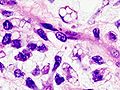
| Image = Dedifferentiated_liposarcoma_-_cropped_-_very_high_mag.jpg | |||
| Width = | |||
| Caption = Liposarcoma. [[H&E stain]]. | |||
| Synonyms = | |||
| Micro = lipoblasts - '''key feature''', chicken wire-like vascular, +/-[[myxoid]] background, cell size variation, thick fibrous septa (important low power feature) | |||
| Subtypes = [[dedifferentiated liposarcoma]], [[myxoid liposarcoma]] (includes round cell liposarcoma), mixed-type liposarcoma, pleomorphic liposarcoma, liposarcoma not otherwise specified (NOS), spindle cell liposarcoma | |||
| LMDDx = [[pleomorphic undifferentiated sarcoma]] | |||
| Stains = | |||
| IHC = S-100 +ve | |||
| EM = | |||
| Molecular = t(12;16)(q13;p11) / TLS-CHOP -- for myxoid liposarcoma | |||
| IF = | |||
| Gross = | |||
| Grossing = | |||
| Site = [[soft tissue lesions|soft tissue]] esp. retroperiteoneum | |||
| Assdx = | |||
| Syndromes = | |||
| Clinicalhx = | |||
| Signs = | |||
| Symptoms = | |||
| Prevalence = most common retroperitoneal sarcoma, uncommon overall | |||
| Bloodwork = | |||
| Rads = thick septae or nodules in a fatty appearing lesion | |||
| Endoscopy = | |||
| Prognosis = poor | |||
| Other = | |||
| ClinDDx = | |||
| Tx = | |||
}} | |||
'''Liposarcoma''' is a [[malignant]] [[Adipocytic tumours|adipocytic tumour]]. | |||
==General== | |||
*Most common malignant sarcoma in the retroperitoneum. | |||
*Not all (large) retroperitoneal adipocytic tumours are liposarcomas. | |||
*Prognosis - dependent on the location. | |||
**Retroperitoneal: poor. | |||
**Extremity: good - disease specific 5-year survival ~ 92%.<ref>{{Cite journal | last1 = Lietman | first1 = SA. | last2 = Barsoum | first2 = WK. | last3 = Goldblum | first3 = JR. | last4 = Marks | first4 = KE. | last5 = Mascha | first5 = E. | last6 = Sundaram | first6 = M. | last7 = Muschler | first7 = G. | title = A 20-year retrospective review of surgically treated liposarcoma at the Cleveland Clinic. | journal = Orthopedics | volume = 30 | issue = 3 | pages = 227-34 | month = Mar | year = 2007 | doi = | PMID = 17375550 }}</ref> | |||
Notes: | |||
*Retroperitoneal sarcomas: #1: liposarcoma, #2: [[pleomorphic undifferentiated sarcoma]], #3: [[leiomyosarcoma]], #4: [[MPNST]]. | |||
**Extremely rare in retroperitoneum: [[synovial sarcoma]]. | |||
==Gross== | |||
*Thick fibrous septa | |||
Radiology:<ref name=pmid9108224/> | |||
*Fibrous septa or nodules.<ref name=pmid16160117>{{Cite journal | last1 = Murphey | first1 = MD. | last2 = Arcara | first2 = LK. | last3 = Fanburg-Smith | first3 = J. | title = From the archives of the AFIP: imaging of musculoskeletal liposarcoma with radiologic-pathologic correlation. | journal = Radiographics | volume = 25 | issue = 5 | pages = 1371-95 | month = | year = | doi = 10.1148/rg.255055106 | PMID = 16160117 }}</ref> | |||
*Myoid liposarcoma - high signal (due to high water content).<ref name=pmid16160117/> | |||
==Microscopic== | |||
Features: | |||
*Lipoblasts - '''key feature'''. | |||
**Large sharply demarcated vacuole. | |||
**Nucleus: | |||
***Hyperchromatic (dark staining) nucleus. | |||
***Eccentric location. | |||
***Nuclear indentation. | |||
*Chicken wire-like vascular. | |||
*+/-[[Myxoid]] background. | |||
*Cell size variation. | |||
*Thick fibrous septa - important low power feature.<ref name=pmid9108224>{{Cite journal | last1 = Hosono | first1 = M. | last2 = Kobayashi | first2 = H. | last3 = Fujimoto | first3 = R. | last4 = Kotoura | first4 = Y. | last5 = Tsuboyama | first5 = T. | last6 = Matsusue | first6 = Y. | last7 = Nakamura | first7 = T. | last8 = Itoh | first8 = T. | last9 = Konishi | first9 = J. | title = Septum-like structures in lipoma and liposarcoma: MR imaging and pathologic correlation. | journal = Skeletal Radiol | volume = 26 | issue = 3 | pages = 150-4 | month = Mar | year = 1997 | doi = | PMID = 9108224 }}</ref> | |||
DDx: | |||
*[[Angiolipoma]]. | |||
*[[Pleomorphic undifferentiated sarcoma]]. | |||
===Images=== | |||
<gallery> | |||
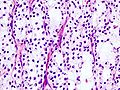
Image: Dedifferentiated liposarcoma - intermed mag.jpg | Dediff. liposar. - shows dediff. component - intermed. mag. (WC) | |||
Image: Dedifferentiated liposarcoma - high mag.jpg | Dediff. liposarc. - high mag. (WC) | |||
Image: Dedifferentiated liposarcoma - very high mag.jpg | Dediff. liposarc. - very high mag. (WC) | |||
Image: Dedifferentiated liposarcoma - cropped - very high mag.jpg | Dediff. liposarc. - lipoblasts - very high mag. (WC) | |||
</gallery> | |||
<gallery> | |||
Image:Myxoid_liposarcoma_%2806%29.JPG | Myxoid liposarcoma. (WC) | |||
Image:Myxoid_liposarcoma_%2805%29.JPG | Myxoid liposarcoma. (WC) | |||
</gallery> | |||
www: | |||
*[http://www.john-libbey-eurotext.fr/e-docs/00/04/09/14/texte_alt_jleejd00046_gr5.jpg Lipoblasts (john-libbey-eurotext.fr)]. | |||
*[http://path.upmc.edu/cases/case302.html Poorly diff. liposarcoma - several images (upmc.edu)]. | |||
===Subtypes=== | |||
Main subtypes:<ref name=Ref_WMSP_601>{{Ref WMSP|601}}</ref> | |||
*Dedifferentiated liposarcoma. | |||
*Myxoid liposarcoma. | |||
**Round cell liposarcoma - a subtype of myxoid liposarcoma<ref name=pmid8554106>{{Cite journal | last1 = Smith | first1 = TA. | last2 = Easley | first2 = KA. | last3 = Goldblum | first3 = JR. | title = Myxoid/round cell liposarcoma of the extremities. A clinicopathologic study of 29 cases with particular attention to extent of round cell liposarcoma. | journal = Am J Surg Pathol | volume = 20 | issue = 2 | pages = 171-80 | month = Feb | year = 1996 | doi = | PMID = 8554106 }} | |||
</ref> that has a worse prognosis;<ref name=pmid21253554>{{Cite journal | last1 = Conyers | first1 = R. | last2 = Young | first2 = S. | last3 = Thomas | first3 = DM. | title = Liposarcoma: molecular genetics and therapeutics. | journal = Sarcoma | volume = 2011 | issue = | pages = 483154 | month = | year = 2011 | doi = 10.1155/2011/483154 | PMID = 21253554 }}</ref> characterized by regions of high cellularity. | |||
*Mixed-type liposarcoma. | |||
*Pleomorphic liposarcoma. | |||
*Liposarcoma not otherwise specified (NOS). | |||
Additional reported type: | |||
*Spindle cell liposarcoma.<ref name=pmid8067512>{{Cite journal | last1 = Dei Tos | first1 = AP. | last2 = Mentzel | first2 = T. | last3 = Newman | first3 = PL. | last4 = Fletcher | first4 = CD. | title = Spindle cell liposarcoma, a hitherto unrecognized variant of liposarcoma. Analysis of six cases. | journal = Am J Surg Pathol | volume = 18 | issue = 9 | pages = 913-21 | month = Sep | year = 1994 | doi = | PMID = 8067512 }}</ref> | |||
====Myxoid liposarcoma==== | |||
*[[AKA]] ''myxoid/round cell liposarcoma''. | |||
*[[AKA]] ''round cell liposarcoma''. | |||
=====Gross===== | |||
Location:<ref name=pmid22052112>{{Cite journal | last1 = Moreau | first1 = LC. | last2 = Turcotte | first2 = R. | last3 = Ferguson | first3 = P. | last4 = Wunder | first4 = J. | last5 = Clarkson | first5 = P. | last6 = Masri | first6 = B. | last7 = Isler | first7 = M. | last8 = Dion | first8 = N. | last9 = Werier | first9 = J. | title = Myxoid\Round Cell Liposarcoma (MRCLS) Revisited: An Analysis of 418 Primarily Managed Cases. | journal = Ann Surg Oncol | volume = 19 | issue = 4 | pages = 1081-1088 | month = Apr | year = 2012 | doi = 10.1245/s10434-011-2127-z | PMID = 22052112 }}</ref> | |||
*90% in lower limb. | |||
*81% deep. | |||
=====Microscopic===== | |||
Features: | |||
*Chickenwire-type blood vessels. | |||
*Clear spaces. | |||
*Adipocytes - may be rare. | |||
DDx: | |||
*[[Low-grade fibromyxoid sarcoma]]. | |||
Images: | |||
*[http://pubs.rsna.org/doi/full/10.1148/radiographics.20.4.g00jl021007 Myxoid liposarcoma (rsna.org)].<ref name=pmid10903690>{{Cite journal | last1 = Sung | first1 = MS. | last2 = Kang | first2 = HS. | last3 = Suh | first3 = JS. | last4 = Lee | first4 = JH. | last5 = Park | first5 = JM. | last6 = Kim | first6 = JY. | last7 = Lee | first7 = HG. | title = Myxoid liposarcoma: appearance at MR imaging with histologic correlation. | journal = Radiographics | volume = 20 | issue = 4 | pages = 1007-19 | month = | year = | doi = 10.1148/radiographics.20.4.g00jl021007 | PMID = 10903690 }}</ref> | |||
*[http://pubs.rsna.org/doi/figure/10.1148/rg.255055106 Myxoid liposarcoma - among other things (rsna.org)].<ref name=pmid16160117>{{Cite journal | last1 = Murphey | first1 = MD. | last2 = Arcara | first2 = LK. | last3 = Fanburg-Smith | first3 = J. | title = From the archives of the AFIP: imaging of musculoskeletal liposarcoma with radiologic-pathologic correlation. | journal = Radiographics | volume = 25 | issue = 5 | pages = 1371-95 | month = | year = | doi = 10.1148/rg.255055106 | PMID = 16160117 }}</ref> | |||
*[http://www.cap.org/apps/cap.portal?_nfpb=true&cntvwrPtlt_actionOverride=%2Fportlets%2FcontentViewer%2Fshow&_windowLabel=cntvwrPtlt&cntvwrPtlt{actionForm.contentReference}=cap_foundation%2FcaseOfMonth%2Ffeb13%2Ffeb_2013_cotm_diagnosis.html&_state=maximized&_pageLabel=cntvwr Myxoid liposarcoma (cap.org)]. | |||
=====Molecular===== | |||
Typically has a [[translocations|translocation]]: | |||
*t(12;16)(q13;p11) TLS-CHOP.<ref name=pmid7805034>{{Cite journal | last1 = Knight | first1 = JC. | last2 = Renwick | first2 = PJ. | last3 = Dal Cin | first3 = P. | last4 = Van den Berghe | first4 = H. | last5 = Fletcher | first5 = CD. | title = Translocation t(12;16)(q13;p11) in myxoid liposarcoma and round cell liposarcoma: molecular and cytogenetic analysis. | journal = Cancer Res | volume = 55 | issue = 1 | pages = 24-7 | month = Jan | year = 1995 | doi = | PMID = 7805034 }}</ref> | |||
*May have the translocation usually seen in [[clear cell sarcoma]]: | |||
*t(12;22)(q13;q12) EWS-ATF1.<ref name=pmid21115923>{{Cite journal | last1 = Suzuki | first1 = K. | last2 = Matsui | first2 = Y. | last3 = Endo | first3 = K. | last4 = Kubo | first4 = T. | last5 = Hasegawa | first5 = T. | last6 = Kimura | first6 = T. | last7 = Ohtani | first7 = O. | last8 = Yasui | first8 = N. | title = Myxoid liposarcoma with EWS-CHOP type 1 fusion gene. | journal = Anticancer Res | volume = 30 | issue = 11 | pages = 4679-83 | month = Nov | year = 2010 | doi = | PMID = 21115923 }}</ref> | |||
====Dedifferentiated liposarcoma==== | |||
*Has an undifferentiated component that, if seen alone, would be diagnosed as [[pleomorphic undifferentiated sarcoma]]. | |||
*The diagnosis depends on the presence of the differentiated component of the tumour, i.e. the presence of lipoblasts. | |||
==IHC== | |||
*MDM2 +ve.<ref name=pmid24227706>{{Cite journal | last1 = Mariño-Enríquez | first1 = A. | last2 = Hornick | first2 = JL. | last3 = Dal Cin | first3 = P. | last4 = Cibas | first4 = ES. | last5 = Qian | first5 = X. | title = Dedifferentiated liposarcoma and pleomorphic liposarcoma: a comparative study of cytomorphology and MDM2/CDK4 expression on fine-needle aspiration. | journal = Cancer Cytopathol | volume = 122 | issue = 2 | pages = 128-37 | month = Feb | year = 2014 | doi = 10.1002/cncy.21362 | PMID = 24227706 }}</ref> | |||
*CDK4 +ve.<ref name=pmid24227706/> | |||
*S-100 +ve ~1/3 of the time. | |||
==See also== | |||
*[[Adipocytic tumours]]. | |||
==References== | |||
{{Reflist|2}} | |||
[[Category:Diagnosis]] | [[Category:Diagnosis]] | ||
[[Category:Adipocytic tumours]] | |||
Revision as of 03:15, 19 September 2014
| Liposarcoma | |
|---|---|
| Diagnosis in short | |
 Liposarcoma. H&E stain. | |
|
| |
| LM | lipoblasts - key feature, chicken wire-like vascular, +/-myxoid background, cell size variation, thick fibrous septa (important low power feature) |
| Subtypes | dedifferentiated liposarcoma, myxoid liposarcoma (includes round cell liposarcoma), mixed-type liposarcoma, pleomorphic liposarcoma, liposarcoma not otherwise specified (NOS), spindle cell liposarcoma |
| LM DDx | pleomorphic undifferentiated sarcoma |
| IHC | S-100 +ve |
| Molecular | t(12;16)(q13;p11) / TLS-CHOP -- for myxoid liposarcoma |
| Site | soft tissue esp. retroperiteoneum |
|
| |
| Prevalence | most common retroperitoneal sarcoma, uncommon overall |
| Radiology | thick septae or nodules in a fatty appearing lesion |
| Prognosis | poor |
Liposarcoma is a malignant adipocytic tumour.
General
- Most common malignant sarcoma in the retroperitoneum.
- Not all (large) retroperitoneal adipocytic tumours are liposarcomas.
- Prognosis - dependent on the location.
- Retroperitoneal: poor.
- Extremity: good - disease specific 5-year survival ~ 92%.[1]
Notes:
- Retroperitoneal sarcomas: #1: liposarcoma, #2: pleomorphic undifferentiated sarcoma, #3: leiomyosarcoma, #4: MPNST.
- Extremely rare in retroperitoneum: synovial sarcoma.
Gross
- Thick fibrous septa
Radiology:[2]
Microscopic
Features:
- Lipoblasts - key feature.
- Large sharply demarcated vacuole.
- Nucleus:
- Hyperchromatic (dark staining) nucleus.
- Eccentric location.
- Nuclear indentation.
- Chicken wire-like vascular.
- +/-Myxoid background.
- Cell size variation.
- Thick fibrous septa - important low power feature.[2]
DDx:
Images
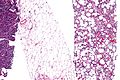
Dediff. liposar. - shows dediff. component - intermed. mag. (WC)
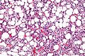
Dediff. liposarc. - high mag. (WC)
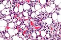
Dediff. liposarc. - very high mag. (WC)
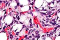
Dediff. liposarc. - lipoblasts - very high mag. (WC)


Myxoid liposarcoma. (WC)
Myxoid liposarcoma. (WC)
.JPG)
.JPG)
www:
Subtypes
Main subtypes:[4]
- Dedifferentiated liposarcoma.
- Myxoid liposarcoma.
- Mixed-type liposarcoma.
- Pleomorphic liposarcoma.
- Liposarcoma not otherwise specified (NOS).
Additional reported type:
- Spindle cell liposarcoma.[7]
Myxoid liposarcoma
Gross
Location:[8]
- 90% in lower limb.
- 81% deep.
Microscopic
Features:
- Chickenwire-type blood vessels.
- Clear spaces.
- Adipocytes - may be rare.
DDx:
Images:
- Myxoid liposarcoma (rsna.org).[9]
- Myxoid liposarcoma - among other things (rsna.org).[3]
- Myxoid liposarcoma (cap.org).
Molecular
Typically has a translocation:
- t(12;16)(q13;p11) TLS-CHOP.[10]
- May have the translocation usually seen in clear cell sarcoma:
- t(12;22)(q13;q12) EWS-ATF1.[11]
Dedifferentiated liposarcoma
- Has an undifferentiated component that, if seen alone, would be diagnosed as pleomorphic undifferentiated sarcoma.
- The diagnosis depends on the presence of the differentiated component of the tumour, i.e. the presence of lipoblasts.
IHC
See also
References
- ↑ Lietman, SA.; Barsoum, WK.; Goldblum, JR.; Marks, KE.; Mascha, E.; Sundaram, M.; Muschler, G. (Mar 2007). "A 20-year retrospective review of surgically treated liposarcoma at the Cleveland Clinic.". Orthopedics 30 (3): 227-34. PMID 17375550.
- ↑ 2.0 2.1 Hosono, M.; Kobayashi, H.; Fujimoto, R.; Kotoura, Y.; Tsuboyama, T.; Matsusue, Y.; Nakamura, T.; Itoh, T. et al. (Mar 1997). "Septum-like structures in lipoma and liposarcoma: MR imaging and pathologic correlation.". Skeletal Radiol 26 (3): 150-4. PMID 9108224.
- ↑ 3.0 3.1 3.2 Murphey, MD.; Arcara, LK.; Fanburg-Smith, J.. "From the archives of the AFIP: imaging of musculoskeletal liposarcoma with radiologic-pathologic correlation.". Radiographics 25 (5): 1371-95. doi:10.1148/rg.255055106. PMID 16160117.
- ↑ Humphrey, Peter A; Dehner, Louis P; Pfeifer, John D (2008). The Washington Manual of Surgical Pathology (1st ed.). Lippincott Williams & Wilkins. pp. 601. ISBN 978-0781765275.
- ↑ Smith, TA.; Easley, KA.; Goldblum, JR. (Feb 1996). "Myxoid/round cell liposarcoma of the extremities. A clinicopathologic study of 29 cases with particular attention to extent of round cell liposarcoma.". Am J Surg Pathol 20 (2): 171-80. PMID 8554106.
- ↑ Conyers, R.; Young, S.; Thomas, DM. (2011). "Liposarcoma: molecular genetics and therapeutics.". Sarcoma 2011: 483154. doi:10.1155/2011/483154. PMID 21253554.
- ↑ Dei Tos, AP.; Mentzel, T.; Newman, PL.; Fletcher, CD. (Sep 1994). "Spindle cell liposarcoma, a hitherto unrecognized variant of liposarcoma. Analysis of six cases.". Am J Surg Pathol 18 (9): 913-21. PMID 8067512.
- ↑ Moreau, LC.; Turcotte, R.; Ferguson, P.; Wunder, J.; Clarkson, P.; Masri, B.; Isler, M.; Dion, N. et al. (Apr 2012). "Myxoid\Round Cell Liposarcoma (MRCLS) Revisited: An Analysis of 418 Primarily Managed Cases.". Ann Surg Oncol 19 (4): 1081-1088. doi:10.1245/s10434-011-2127-z. PMID 22052112.
- ↑ Sung, MS.; Kang, HS.; Suh, JS.; Lee, JH.; Park, JM.; Kim, JY.; Lee, HG.. "Myxoid liposarcoma: appearance at MR imaging with histologic correlation.". Radiographics 20 (4): 1007-19. doi:10.1148/radiographics.20.4.g00jl021007. PMID 10903690.
- ↑ Knight, JC.; Renwick, PJ.; Dal Cin, P.; Van den Berghe, H.; Fletcher, CD. (Jan 1995). "Translocation t(12;16)(q13;p11) in myxoid liposarcoma and round cell liposarcoma: molecular and cytogenetic analysis.". Cancer Res 55 (1): 24-7. PMID 7805034.
- ↑ Suzuki, K.; Matsui, Y.; Endo, K.; Kubo, T.; Hasegawa, T.; Kimura, T.; Ohtani, O.; Yasui, N. (Nov 2010). "Myxoid liposarcoma with EWS-CHOP type 1 fusion gene.". Anticancer Res 30 (11): 4679-83. PMID 21115923.
- ↑ 12.0 12.1 Mariño-Enríquez, A.; Hornick, JL.; Dal Cin, P.; Cibas, ES.; Qian, X. (Feb 2014). "Dedifferentiated liposarcoma and pleomorphic liposarcoma: a comparative study of cytomorphology and MDM2/CDK4 expression on fine-needle aspiration.". Cancer Cytopathol 122 (2): 128-37. doi:10.1002/cncy.21362. PMID 24227706.