Lipoma
Jump to navigation
Jump to search
The printable version is no longer supported and may have rendering errors. Please update your browser bookmarks and please use the default browser print function instead.
| Lipoma | |
|---|---|
| Diagnosis in short | |
 Mature adipose tissue (lipoma). H&E stain. | |
|
| |
| Synonyms | steatoma (old term, ambiguous) |
|
| |
| LM | mature adipocytes |
| LM DDx | liposarcoma, benign fat |
| IHC | S-100 +ve |
| Molecular | MDM2/CDK4 amplification absent |
| Gross | soft yellow tissue - typically with a thin capsule and lobulated |
| Site | soft tissue |
|
| |
| Signs | pillow sign (endoscopy) |
| Prevalence | common |
| Endoscopy | smooth yellow coloured submucosal lesion |
| Prognosis | benign |
| Treatment | surgical removal or follow-up |
Lipoma is a benign adipocytic tumour.
General
- Benign.
- Several variants exist.
- Angiolipoma - one of the (classically) painful skin lesions.
- May be seen in association with MERRF syndrome (myoclonic epilepsy with ragged-red fibres).[1]
- May be seen in the context of Madelung's disease.[2]
Gross
- Soft yellow tissue - typically lobulated and with a very thin capsule.
Note:
- May be quite large ~10 cm.
- Thigh lesions are more likely to the malignant than other sites.[3]
Microscopic
Features:
- Collection of mature adipocytes.
- Variation of size may be seen -- should prompt a search for lipoblasts.[4]
Notes:
- Microscopically not definitely distinguishable from mature clump of fat.
- The lesion must be labeled lipoma (by the clinican) to be signed-out as such.
DDx:
- Liposarcoma - increased number of blood vessels,[5] esp. chickenwire-like vessels, fibrous septae.
- Benign adipose tissue.
Images:
Variants
Angiolipoma
Microscopic:
- Numerous blood vessels present.
- +/-Microthrombi.
DDx:
Myolipoma
General:
Microscopic:[8]
- Mature adipose tissue.
- Benign smooth muscle - usually ~ 2x amount of fat.
Note:
- If skeletal muscle is present consider intramuscular lipoma.[10]
IHC:[8]
- Actin +ve.
- Desmin +ve.
Images:
Images
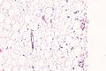
Lipoma - intermed. mag.
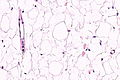
Lipoma - high mag.
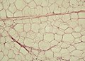
Mature fat. (WC)


Molecular
- MDM2/CDK4 gene amplification absent.
- Testing suggested in lesions greater than 10 cm, thigh lesions and lesions with cytologic atypia.[3]
Sign out
Large lesion looks like lipoma
Bland lesions may be well-differentiated liposarcoma.[12] Lesions >10 cm should be of concern.
Lesion (Submitted as "Lipoma"), Right Neck, Excision:
	- Bland appearing adipose tissue suggestive of lipoma, see comment.
	- One benign lymph node.

Comment:
Due to the size of the lesion, the case will be sent to a soft tissue pathologist for review.
Block letter
SUBCUTANEOUS TISSUE ("LIPOMA"), LEFT AXILLA, EXCISION:
- MATURE ADIPOSE TISSUE CONSISTENT WITH LIPOMA.
LESION ("LIPOMA"), SPERMATIC CORD (LATERALITY NOT SPECIFIED), EXCISION:
- MATURE ADIPOSE TISSUE CONSISTENT WITH LIPOMA.
Colonic lipoma (clinically suspected)
B. SIGMOID COLON AT 55 CM, BIOPSY:
- COLORECTAL-TYPE MUCOSA WITHIN NORMAL LIMITS WITH A SMALL AMOUNT OF SUBMUCOSAL
  ADIPOSE TISSUE; COMPATIBLE WITH CLINICAL IMPRESSION OF LIPOMA.
Mirco
The sections show mature adipocytes. There is no increase in vascularity. No thick fibrous septa are present.
See also
References
- ↑ Jones, AP.; Lewis, CJ.; Dildey, P.; Hide, G.; Ragbir, M. (Jan 2012). "Lipoma or liposarcoma? A cautionary case report.". J Plast Reconstr Aesthet Surg 65 (1): e11-4. doi:10.1016/j.bjps.2011.08.004. PMID 21865105.
- ↑ Mayo Yáñez, M.; González Poggioli, N.; Álvarez-Buylla Blanco, M.; Herranz González-Botas, J. (Nov 2017). "Benign symmetric lipomatosis with lingual involvement: Case report and literature review.". J Stomatol Oral Maxillofac Surg. doi:10.1016/j.jormas.2017.11.006. PMID 29129710.
- ↑ 3.0 3.1 Wong, DD.; Low, IC.; Peverall, J.; Robbins, PD.; Spagnolo, DV.; Nairn, R.; Carey-Smith, RL.; Wood, D. (Apr 2016). "MDM2/CDK4 gene amplification in large/deep-seated 'lipomas': incidence, predictors and clinical significance.". Pathology 48 (3): 203-9. doi:10.1016/j.pathol.2016.02.007. PMID 27020493.
- ↑ Mentzel, T.; Fletcher, CD. (1995). "Lipomatous tumours of soft tissues: an update.". Virchows Arch 427 (4): 353-63. PMID 8548119.
- ↑ Yang, YJ.; Damron, TA.; Cohen, H.; Hojnowski, L. (Oct 2001). "Distinction of well-differentiated liposarcoma from lipoma in two patients with multiple well-differentiated fatty masses.". Skeletal Radiol 30 (10): 584-9. doi:10.1007/s002560100395. PMID 11685482.
- ↑ Friedberg, MK.; Chang, IL.; Silverman, NH.; Ramamoorthy, C.; Chan, FP. (May 2006). "Images in cardiovascular medicine. Near sudden death from cardiac lipoma in an adolescent.". Circulation 113 (21): e778-9. doi:10.1161/CIRCULATIONAHA.105.589630. PMID 16735681. http://circ.ahajournals.org/content/113/21/e778.full.
- ↑ URL: http://www.webmedcentral.com/article_view/1878. Accessed on: 14 March 2013.
- ↑ 8.0 8.1 8.2 Murphey, MD.; Carroll, JF.; Flemming, DJ.; Pope, TL.; Gannon, FH.; Kransdorf, MJ.. "From the archives of the AFIP: benign musculoskeletal lipomatous lesions.". Radiographics 24 (5): 1433-66. doi:10.1148/rg.245045120. PMID 15371618. http://radiographics.rsna.org/content/24/5/1433.long.
- ↑ Meis, JM.; Enzinger, FM. (Feb 1991). "Myolipoma of soft tissue.". Am J Surg Pathol 15 (2): 121-5. PMID 1703396.
- ↑ URL: http://surgpathcriteria.stanford.edu/softfat/lipoma/intramuscular_lipoma.html. Accessed on: 14 March 2013.
- ↑ Lee, YS.; Park, SE.; Lee, JU.; Choi, ES.. "MRI of a subcutaneous myolipoma in the ankle: a case report.". Korean J Radiol 12 (5): 641-5. doi:10.3348/kjr.2011.12.5.641. PMC 3168809. PMID 21927569. https://www.ncbi.nlm.nih.gov/pmc/articles/PMC3168809/.
- ↑ URL: https://www.ncbi.nlm.nih.gov/pmc/articles/PMC3422587/. Accessed on: 3 June 2017.