Leydig cell hyperplasia
Jump to navigation
Jump to search
The printable version is no longer supported and may have rendering errors. Please update your browser bookmarks and please use the default browser print function instead.
| Leydig cell hyperplasia | |
|---|---|
| Diagnosis in short | |
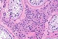
 Leydig cell hyperplasia. H&E stain. (WC) | |
|
| |
| LM | abundant Leydig cells interspersed between seminiferous tubules with only Sertoli cells, Leydig cells do not displace or compress the seminiferous tubules |
| LM DDx | Leydig cell tumour, testicular adrenal rest tumour |
| Site | testis, ovary |
|
| |
| Prevalence | rare |
| Radiology | hypoechoic lesions on ultrasound, often multiple |
| Prognosis | benign |
Leydig cell hyperplasia is an uncommon benign pathology of the testis.[1] It may be seen in the ovary.
General
- Benign.
- Uncommonly seen in isolation by pathology.
A longer list of causes:[2]
- Congenital (primary).
- Secondary causes:
- Germ cell loss/decrease:
- Cryptorchidism.
- Advanced age.
- Endocrine-related:
- Adrenal hyperplasia.
- Excess hCG - exogenous or tumour.
- Germ cell loss/decrease:
Other associations:[2]
- Klinefelter's syndrome.[3]
- Pernicious anemia.
- Alcoholism.
- Infection - syphilis, tuberculosis.
Gross
Features:
- Unremarkable gross appearance of parenchyma. (???)
- Normal or small testis. (???)
Note:
- Hypoechoic lesions on ultrasound - often multiple, small.[3]
Microscopic
Features:
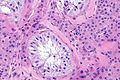
- Abundant Leydig cells interspersed between seminiferous tubules with only Sertoli cells.
- Leydig cells do not displace or compress the seminiferous tubules.
Note:
- May form nodules up to 6 mm.[1]
DDx:
Images
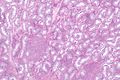
LCH - very low mag. (WC)
LCH - very low mag. (WC)
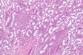
LCH - low mag. (WC)
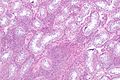
LCH - intermed. mag. (WC)
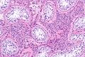
LCH - high mag. (WC)
LCH - high mag. (WC)


Sign out
Testicle and Cord, Right, Orchitectomy:
- Leydig cell hyperplasia.
- Atrophic testis.
- NEGATIVE for germ cell neoplasia in situ (intratubular germ cell neoplasia).
- NEGATIVE for malignancy.

Comment:
Immunostains confirm the morphologic impression.

The Leydig cells are POSITIVE for inhibin, calretinin and melan A. The section is
NEGATIVE for PLAP and has a benign pattern for D2-40.
See also
References
- ↑ 1.0 1.1 Carucci, LR.; Tirkes, AT.; Pretorius, ES.; Genega, EM.; Weinstein, SP. (Feb 2003). "Testicular Leydig's cell hyperplasia: MR imaging and sonographic findings.". AJR Am J Roentgenol 180 (2): 501-3. doi:10.2214/ajr.180.2.1800501. PMID 12540460.
- ↑ 2.0 2.1 Naughton, CK.; Nadler, RB.; Basler, JW.; Humphrey, PA. (Feb 1998). "Leydig cell hyperplasia.". Br J Urol 81 (2): 282-9. doi:10.1046/j.1464-410X.1998.00503.x. PMID 9488073.
- ↑ 3.0 3.1 Sterbis, J.; E-Nunu, T. (2015). "Leydig cell hyperplasia in the setting of Klinefelter syndrome.". BMJ Case Rep 2015. doi:10.1136/bcr-2015-209805. PMID 26209412.