Intradermal nevus
| Intradermal nevus | |
|---|---|
| Diagnosis in short | |
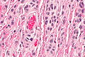
 Intradermal nevus. H&E stain. | |
|
| |
| Synonyms | intradermal melanocytic nevus |
|
| |
| LM | nests of melanocytes in dermis (only), melanocytes "mature" with depth, usu. no mitoses (occ. superficial), no destruction of surrounding structures, no conspicuous nucleoli, no significant melanocyte enlargement |
| LM DDx | malignant melanoma (nevoid), junctional nevus, compound nevus, dysplastic nevus, skin tag, mastocytosis |
| Gross | pigment skin lesion, usu. small, regular border, no irregularity in pigmentation |
| Site | skin - see melanocytic lesions and common nevus |
|
| |
| Prevalence | very common |
| Prognosis | benign |
| Clin. DDx | pigmented skin lesions |
| Treatment | none required, may be excised for cosmetic reasons |
Intradermal nevus (abbreviated IDN), also intradermal melanocytic nevus, is a common benign melanocytic lesion.
The intradermal nevus is in the large group common nevus. In common language, nevus is known as a mole.
In the oral cavity these are known as intramucosal nevi.
General
- Benign.
- Common.
- Think melanoma.
Clinical:
- ABCD = asymmetric, borders (irregular), colour (black), diameter (large).
Microscopic
Features:
- Symmetrical lesion.
- "Matures" with depth.
- Less cellular with depth.
- Less nuclear atypia with depth.
- Smaller cells with depth.
- Smaller nests with depth.
- Rare mitoses (superficial).
- No deep mitoses.
- No destruction of surrounding structures.
- No nucleoli.
- In the dermis only - key feature.
- +/-Adipocytes - uncommon.[1]
DDx:
- Malignant melanoma (nevoid).
- Dysplastic nevus.
- Junctional nevus.
- Compound nevus.
- Skin tag.
- Mastocytosis.
Images
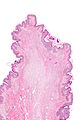
IDN - very low mag.
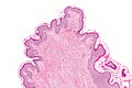
IDN - low mag.
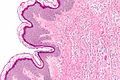
IDN - intermed. mag.
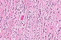
IDN - high mag.
IDN - very high mag.


Sign out
Skin, Left Alar Crease, Biopsy:
- Benign intradermal nevus.
Block letters
SKIN LESION, BACK, PUNCH BIOPSY:
- BENIGN INTRADERMAL NEVUS.
Adipocytes present
SKIN LESION, BACK, PUNCH BIOPSY:
- BENIGN INTRADERMAL NEVUS WITH MATURE ADIPOCYTES.
Clinically suspicious
SKIN LESION, LEFT UPPER ARM, PUNCH BIOPSY:
- BENIGN INTRADERMAL NEVUS, INCOMPLETELY EXCISED.

COMMENT:
HMB-45 marks very rare superficial cells. Ki-67 staining does not show apparent melanocyte
staining. MITF marks the melanocytes and demonstrates maturation of the melanocytes with depth.
Micro
The sections show melanocytes in the dermis separated from the epidermis by a Grenz zone. The lesion is symmetrical in its architecture and pigment distribution. Superficially, melanocytes are in nests. Melanocytes mature with depth. No mitotic activity is appreciated.
The lesion is completely excised in the plane of section.
IDN with congenital features
The sections show melanocytes in the dermis separated from the epidermis by a Grenz zone. The lesion is symmetrical in its architecture. Superficially, melanocytes are in nests. Melanocytes mature with depth and track along adnexal structures. No mitotic activity is appreciated. The lesion is incompletely excised in the plane of section.