Hepatitis B
Hepatitis B is a relatively common medical liver disease caused by the hepatitis B virus.
General
- May lead to hepatocellular carcinoma without cirrhosis.
- High prevalence.
- Diagnosis is by serology - details of serologic testing are in the medical liver disease article.
- A vaccination is available and done routinely in a many jurisdictions.[1]
Associated pathology:
Microscopic
Features:
- Lobular inflammation - this is non-specific finding.
- Hepatocyte necrosis:
- Necrotic hepatocytes look a lot like neutrophils - however:
- Cytoplasm is more pink.
- Round apoptotic bodies.
- Necrotic hepatocytes look a lot like neutrophils - however:
- Hepatocyte necrosis:
- Ground glass hepatocytes - important.
DDx:
Image

GGH - high mag. (WC)


42 yo woman with bepatitis B virus, modified HAI necroinflammatory grade 5, modified HAI fibrosis stage 1. (would be Metavir activity index 1, fibrosis stage 1). A Triads (red arrows) are slightly cellular, without much interface inflammation, with occasional spots suggestive of spotty necrosis (black arrows). B. Triads, this one being the most expanded, show a mild infiltrate (HAI score 1) of lymphocytes and macrophages, with only a small amount present at the interface between triad and lobule. C. A maximum of two foci of spotty necrosis (arrows) were seen in a 10X field (HAI score 2). D. Occasional foci of zone 3 collapse (thick black lines, green arrows), HAI score 1, and piecemeal necrosis (blue arrows) HAI score 1, were seen. E. Trichrome shows portal fibrosis of most triads, but no bridges, or nodules (HAI stage 2).
A. 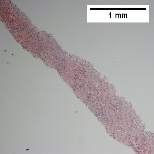 B.
B. 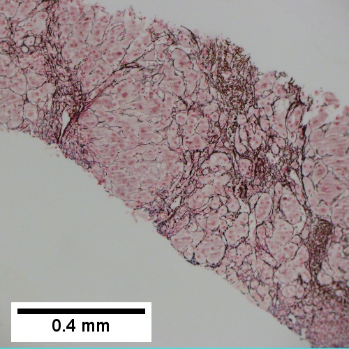
C. 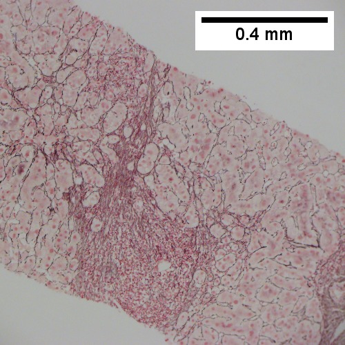 D.
D. 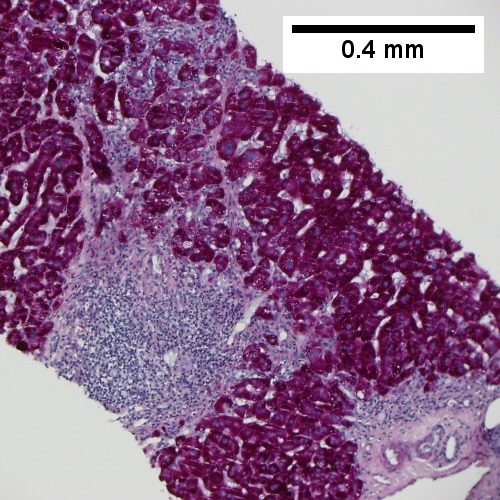
E. 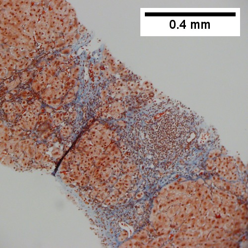 F.
F. 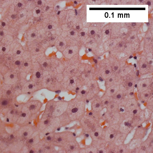
Hepatitis B. Metavir activity index 3. Piecemeal necrosis 2. Lobular necrosis 2. Fibrosis stage 3. A. Portal triads, expanded, inflamed and without sharp edges. Inflammation amid hepatocytes. B. Reticulin with bridging necrosis. C. Reticulin with extensive piecemeal necrosis. D. PAS without diastase with extensive piecemeal necrosis. E. Trichrome with bridging, no nodules or extensive bridging on slides as a whole. F. Ground glass hepatocytes.
A. ![Hepatitis B virus. Metavir activity index 3 {PMN 2 LN 2]. Metavir fibrosis stage 4 (advanced fibrosis/cirrhosis).](/w/images/thumb/0/05/1_HBV_2_680x512px.tif/lossy-page1-680px-1_HBV_2_680x512px.tif.jpg) B.
B. ![Hepatitis B virus. Metavir activity index 3 {PMN 2 LN 2]. Metavir fibrosis stage 4 (advanced fibrosis/cirrhosis).](/w/images/thumb/0/09/2_HBV_2_680x512px.tif/lossy-page1-680px-2_HBV_2_680x512px.tif.jpg)
C. ![Hepatitis B virus. Metavir activity index 3 {PMN 2 LN 2]. Metavir fibrosis stage 4 (advanced fibrosis/cirrhosis).](/w/images/thumb/a/a3/3_HBV_2_680x512px.tif/lossy-page1-680px-3_HBV_2_680x512px.tif.jpg) D.
D. ![Hepatitis B virus. Metavir activity index 3 {PMN 2 LN 2]. Metavir fibrosis stage 4 (advanced fibrosis/cirrhosis).](/w/images/thumb/2/2a/4_HBV_2_680x512px.tif/lossy-page1-680px-4_HBV_2_680x512px.tif.jpg)
E. ![Hepatitis B virus. Metavir activity index 3 {PMN 2 LN 2]. Metavir fibrosis stage 4 (advanced fibrosis/cirrhosis).](/w/images/thumb/1/1b/5_HBV_2_680x512px.tif/lossy-page1-680px-5_HBV_2_680x512px.tif.jpg) F.
F. ![Hepatitis B virus. Metavir activity index 3 {PMN 2 LN 2]. Metavir fibrosis stage 4 (advanced fibrosis/cirrhosis).](/w/images/thumb/7/73/6_HBV_2_680x512px.tif/lossy-page1-680px-6_HBV_2_680x512px.tif.jpg)
Hepatitis B virus. Metavir activity index 3 {PMN 2 LN 2]. Metavir fibrosis stage 4 (advanced fibrosis/cirrhosis). A. A fragmented specimen shows apparent nodules [black arrows]. B. Inflammatory cells extending from band suggest piecemeal necrosis [green arrows]; inflammatory foci apart from band denote spotty necrosis [blue arrows]. C. Reticulin stain shows piecemeal necrosis as black lines about hepatocyte clusters at band [blue arrows], regeneration as more than one cell thick cords [green arrows] and hepatocyte rosettes [magenta arrows]. D. On PAS without diastase, piecemeal necrosis can be seen as pink hepatocyte cytoplasmic fragments amid inflammatory cells [arrows]. E. The nodules seen at low power are confirmed to be collagen bounded, but the appreciation can be made frustrated by intense inflammation. F. Examination elsewhere shows a definite nodule pair amid fibrosis [green arrows]; note on the right the fibrous band extending from the portal triad [black arrow] with sinusoids being far more often perpendicular to the triad than they were in the regenerative nodules to the surrounding bands.
IHC
- Hepatitis B +ve.
See also
References
- ↑ Leuridan, E.; Van Damme, P. (Jul 2011). "Hepatitis B and the need for a booster dose.". Clin Infect Dis 53 (1): 68-75. doi:10.1093/cid/cir270. PMID 21653306.