Hale's colloidal iron stain
Jump to navigation
Jump to search
The printable version is no longer supported and may have rendering errors. Please update your browser bookmarks and please use the default browser print function instead.
Use
- Chromophobe renal cell carcinoma vs. renal oncocytoma - chromophobe renal cell carcinoma +ve.[1]
Interpretation
- Blue (granular cytoplasmic) staining is positive.[2]
Images:
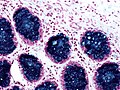
Hale's colloidal iron staining - high mag. (WC)

Notes:
- Often described as a "fastidious" (difficult/demanding) stain.[3]
- A few staff think this is a totally useless stain.[4]
- A variant exists known as the Muller and Mowry modification of Hale's colloidal iron stain (AKA Müller-Mowry stain).[5]
See also
References
- ↑ Tickoo SK, Amin MB, Zarbo RJ (April 1998). "Colloidal iron staining in renal epithelial neoplasms, including chromophobe renal cell carcinoma: emphasis on technique and patterns of staining". Am. J. Surg. Pathol. 22 (4): 419–24. PMID 9537468. http://meta.wkhealth.com/pt/pt-core/template-journal/lwwgateway/media/landingpage.htm?issn=0147-5185&volume=22&issue=4&spage=419.
- ↑ Humphrey, Peter A; Dehner, Louis P; Pfeifer, John D (2008). The Washington Manual of Surgical Pathology (1st ed.). Lippincott Williams & Wilkins. pp. 682. ISBN 978-0781765275.
- ↑ URL: http://www.merriam-webster.com/dictionary/fastidious?show=0&t=1319550566. Accessed on: 25 October 2011.
- ↑ ALS. On several occasions in 2009.
- ↑ Mete, O.; Kilicaslan, I.; Gulluoglu, MG.; Uysal, V. (Dec 2005). "Can renal oncocytoma be differentiated from its renal mimics? The utility of anti-mitochondrial, caveolin 1, CD63 and cytokeratin 14 antibodies in the differential diagnosis.". Virchows Arch 447 (6): 938-46. doi:10.1007/s00428-005-0048-6. PMID 16133362.