Hale's colloidal iron stain
Jump to navigation
Jump to search
Use
- Chromophobe renal cell carcinoma vs. renal oncocytoma - chromophobe renal cell carcinoma +ve.[1]
Notes:
Interpretation
- Blue (granular cytoplasmic) staining is positive.[2]
Images:
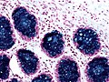
Hale's colloidal iron staining - high mag. (WC)

Notes:
- Often described as a "fastidious" (difficult/demanding) stain.[3]
- A few staff think this is a totally useless stain.[4]
- A variant exists known as the Muller and Mowry modification of Hale's colloidal iron stain (AKA Müller-Mowry stain).[5]
- ↑ Tickoo SK, Amin MB, Zarbo RJ (April 1998). "Colloidal iron staining in renal epithelial neoplasms, including chromophobe renal cell carcinoma: emphasis on technique and patterns of staining". Am. J. Surg. Pathol. 22 (4): 419–24. PMID 9537468. http://meta.wkhealth.com/pt/pt-core/template-journal/lwwgateway/media/landingpage.htm?issn=0147-5185&volume=22&issue=4&spage=419.
- ↑ Humphrey, Peter A; Dehner, Louis P; Pfeifer, John D (2008). The Washington Manual of Surgical Pathology (1st ed.). Lippincott Williams & Wilkins. pp. 682. ISBN 978-0781765275.
- ↑ URL: http://www.merriam-webster.com/dictionary/fastidious?show=0&t=1319550566. Accessed on: 25 October 2011.
- ↑ ALS. On several occasions in 2009.
- ↑ Mete, O.; Kilicaslan, I.; Gulluoglu, MG.; Uysal, V. (Dec 2005). "Can renal oncocytoma be differentiated from its renal mimics? The utility of anti-mitochondrial, caveolin 1, CD63 and cytokeratin 14 antibodies in the differential diagnosis.". Virchows Arch 447 (6): 938-46. doi:10.1007/s00428-005-0048-6. PMID 16133362.