Gynecologic cytopathology
Gynecologic cytopathology is a subset of cytopathology. Gynecologic usually refers to Pap test specimens, i.e. uterine cervix, vaginal vault; other gynecologic specimens are considered non-gynecologic.
This article deals only with cervical cytopathology. An introduction to cytopathology is in the cytopathology article.
Preparation
The standard for Pap test is the Papanicolaou stain. It is described in the staining article and discussed in the context of cytopathology in the cytopathology article.
Slide marking conventions
Conventions are important for facilitating communication between various team members. They are discussed in the cytopathology article.
Normal cells
Squamous cell types:[1]
- Intermediate cells:
- In clusters or single.
- 30-50 micrometres in diameter.
- Associated with progesterone - (light) blue.
- This is the cell of reference in Pap test, i.e. other cells are measured against this cell when assessing a Pap test.
- Parabasal cells:
- Blue-grey.
- Assoc. with atrophy.
- Basal cells:
- Small cells.
- Rarely seen.
- Superficial cell:[2]
- Nucleus smaller than for intermediate cell.
- Cytoplasm red.
- Orange staining superficial cells are hypermature - suggests (abnormal) keratinization.
Glandular cells:[3]
- Sheets of cells with regular spacing.
- Relatively high NC ratio (when compared to intermediate cells).
- Nucleoli (like most glandular cells).
- Nucleus approximately the size of an intermediate cell nucleus.
Mix of cells
The mix of cells is dependent on age and hormones:[4]
- Progesterone - makes the Pap test blue... more intermediate cells.
- Yonger patients have a mix of cells.
- Menopausal patients... more parabasal cells.
- Older patients... more estrogen, glycogen.
Abnormal non-malignant cells
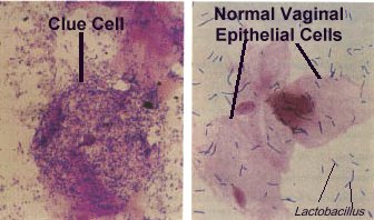
- Clue cells:
- Squamous metaplasia:
- "Dense" cytoplasm.
- Nucleus ~2X the size of an intermediate cell nucleus.
- Nucleoli.
- Note:
- Squamous metaplastic cells have a similar appearance to parabasal cells; they cannot be differentiated on morphologic grounds.
- Squamous metaplastic cells have a high NC ratio - they are differentiated from HSIL via nuclear features (dark staining + irregular nuclear contour = HSIL).
- Slight nuc. contour irregularies are accepted, may be darker staining.
- Endometrial cells:[7]
- Sheet with well-defined border that is bilayered, i.e. clump of epithelioid cells surrounded by spindle cells.
- Scant cytoplasm.
- Chromatin clumping.
- Raisin-like nuclei - approximately the size of an intermediate cell nucleus.
- Nuclei can be considered normal if nucleus less than 2X the size of an intermediate cell nucleus.
- Notes:
- Endometrial cells may appear irregular in the context of an intrauterine device (IUD); abnormalities in the context of an IUD are often ignored.
- The presence of endometrial cells on a Pap test on a woman >=40 years old (per Bethesda guidelines) should be noted in the pathology report[8] - this prompts an endometrial biopsy.
- In my humble opinion, reporting benign endometrial cells in premenopausal women is not evidence-based practise; the practise is driven by lawsuit-paranoia in the USA.
- Atrophy:[9]
- Cells smaller.
- Cytoplasm grey/blue.
- No "dancing"/"sparkling" chromatin.
- +/-"Dirty" background - degenerated cells, inflammatory cells (neutrophils, histiocytes).
- May mimic "dirty" background of tumour, i.e. 'tumour diathesis'.
- Note:
- Usually older women.
- Main DDx is HSIL which has chromatin changes.
Images:
Glycogen halos versus HPV effect
| HPV effect (koilocyte) | Glycogen halo | |
|---|---|---|
| Discolouration of halo | Clear | Yellow |
| Nuclear changes | Associated with nuc. changes | Normal nuclei |
| Cell-to-cell variability | No - all clear | Yes - some yellow some clear |
Gynecologic pathology in tables
Normal cells
| Cell | Architecture | Cell borders | Cytoplasm | DNA | DDx |
|---|---|---|---|---|---|
| Intermediate cell (IC) | Single cells | Irregular | Blue, abundant | Small nucleus (~ size of PMN), no nucleolus | - |
| Superficial cell (SC) | Single cells | Irregular | Red, abundant | Small nucleus, 1/2 size of IC nucleus, no nucleolus | - |
| Squamous metaplastic cell | Single cells/clumps of cells | Smooth/oviod shape | Dense, dark blue | 2X IC nucleus, nucleolus, no membrane irreg., no chromatin changes | DDx: HSIL, basal cell |
| Endometrial cell | Well-circumscribed clump/ball of cells with squamoid covering cells | Indistinct within cluster | Blue, small/very scant | Small, dark, nuclear moulding, degenerative changes (chromatin clumping) | DDx: HSIL, basal cell. |
| Glandular (endocervical) cell | Sheets of cells with regular spacing, columnar morphology may be apparent, +/-pallisading at edge of clump | Often distict | Blue, scant-to-moderate | Nucleus ~ size of an IC nucleus, no membrane irreg., no chromatin changes | DDx: endometrial cell |
| Atrophy | Single cells/groups | Well-circumscribed | Grey/blue dense, may be scant | Large NC ratio, nuc. membrane irregularities, NO chromatin clumping[10] | DDx: HSIL |
Abnormal cells
| Cell | Architecture | Cell borders | Cytoplasm | DNA | Other |
|---|---|---|---|---|---|
| Low-grade squamous intraepithelial lesion (LSIL) | Single cells/groups | Irregular or moderately-circumscribed | Blue, abundant - NC ratio ~ 1:3 | Large nucleus (3-4X IC nuc. - see Note 1), perinuclear clearing, nuc. membrane irregularities, chromatin clumping | DDx: HSIL, reactive changes |
| High-grade squamous intraepithelial lesion (HSIL) | Single cells/groups | Well-circumscribed | Dark blue, scant - NC ratio ~ 1:2 | Large nucleus (3-4X IC nuc. - see Note 1), nuc. membrane irregularities, clumping of coarse chromatin, dark nuc. staining, +/- small nucleoli | DDx: squamous metaplasia, atrophy with atypia |
| Atypical glandular cell (AGC) | Single cells/groups | Usually well-circumscribed (?) | Dark blue dense, scant | Moderately enlarged nucleus (~2X IC nuc.), nuc. membrane irregularities, chromatin clumping, dark nuc. staining, nucleoli | DDx: AIS, HSIL |
| Adenocarinoma in situ (AIS) | Single cells/groups | Usually well-circumscribed | Dark blue dense, scant | Large nucleus (>=2X IC nuc.), nuc. membrane irregularities, chromatin clumping, dark nuc. staining, nucleoli (very common), pseudostratification (as in endocervical AIS) | DDx: AGC, HSIL |
| Features of SCC (see Note 2) | Large clusters of cells with irreg. edge and "streaming", +/-blood, necrotic debris | Poorly seen | Dark blue dense, scant | Large NC ratio, nucleolus, nuc. membrane irregularities, chromatin clumping | DDx: HSIL |
Note 1:
- LSIL/HSIL nucleus - at least 3X IC nucleus.
- ASCUS nucleus - at least 2.5X IC nucleus.
- 3X is not an absolute requirement to call SIL, i.e. SIL may be called with a smaller nucleus in circumstances where other nuclear features are at the extremus of malignant.
- ASCUS nucleus - at least 2.5X IC nucleus.
- Large nuclear size, membrane irregularities, "clumpy" chromatin and dark nuc. staining - are the key features.
- Perinuclear clearing is quite subjective.
- The best perinuclear halos have a sharp punched-out edge.
- Perinuclear clearing is quite subjective.
Note 2:
- By definition, it is not possible to diagnose squamous cell carcinoma (SCC) on a pap test as one cannot demonstrate stromal invasion.
HSIL versus LSIL
| HSIL | LSIL | |
|---|---|---|
| NC ratio[11] - see Note 1 | ~1:2 | ~1:3 |
| Nuclear membrane irregularities | Marked - distinct notches | Moderate |
| Chromatin granularity | Coarse, clumped, +/-nucleolus (red) | Coarse, no nucleolus |
| Cytoplasmic staining | Dark | Light |
| Perinuclear clearing | Usually absent | Often present |
| Binucleation | Uncommon | May be present |
| Maturity of squamous cell | Normal maturity | Hypermature (orangeophilic cell present) |
| Image (example) | HSIL (WC) | LSIL (WC), LSIL & endoCx (WC) |
Note 1:
- The single most useful feature is NC ratio but it is not definitive; NC ratio should be evaluated in the context of nuclear irregularities (nuclear membrane smoothness, chromatin pattern, presence of nucleolus).[11]
- It may be easier to think in terms of cell size - approximate values are:
- HSIL cells: < 1/2 size of IC.
- LSIL cells: classically the size of IC.
Infectious organisms
| Disease | Organism | Group | Dx features | Associated features | Clinical | Reference | Image |
|---|---|---|---|---|---|---|---|
| Trichomoniasis | Trichomonas vaginalis | Protozoan | Pale-grey fluffy cytoplasm with well-defined nucleus, approx. 30 micrometres. | Acute inflammation (PMNs) | Sexually transmitted | [12] | T. vaginalis - Pap stain (WC), Trichomonas - Pap stain (WC) |
| Candidiasis | Candida albicans | Fungi | Branching hyphae ~= 1/2 the dia. of IC nucleus, red | PMNs | ? | ? | Candida on Pap test (WC) |
| Herpes | Herpes simplex virus (HSV 1 - less commonly, HSV 2 - more commonly) | Virus | Large ground glass nuclei then multinucleation with moulding & inclusions with clear halo | ? | Sexually transmitted | ? | HSV (WC),HSV (WC), Herpes simplex virus - surgical (virology.org) |
| Actinomycetes | Actinomycetes | Gram-positive bacteria | Clusters of cocci in chains - hyphae-like appearance | ? | Should prompt removal of IUD, if present. | [13] | Actinomycetes (upenn.edu) |
| Bacterial vaginosis (see Note 1) | Gardnerella vaginalis | Gram-variable rod | "Clue cell": bacterial clusters attached to a purple squamous cell | ? Assoc. | Fishy smell | ? | Bacterial vaginosis (WC), Clue cell (atsu.edu) |
Note 1:
- Usually not reported.
Adequacy of specimens
There is a generally accepted standard for cervical (liquid-based) cytology specimens:[14]
- >5000 squamous cells/slide, if no abnormality is present.
- If abnormal cells are present, any number of cells is acceptable.
- This works-out to approx. 4 cells/HPF.
- Where: HPF = area seen at 400X with an eye piece diameter is ~22 mm.
- 10 HPFs are counted and a table is used to see whether the sample is adequate.
- This works-out to approx. 4 cells/HPF.
- If abnormal cells are present, any number of cells is acceptable.
Note:
- The standard for conventional pap smears is: 8000-12000 squamous cells/slide.[15]
Transformation zone (TZ)
The presence of the TZ should be commented on:[16]
- An adequate TZ is 10 cells - endocervical cells or squamous metaplastic cells (per Bethesda).
Difficulties in obtaining a TZ may arise in the following populations:
- Pregnant (endocervical canal not sampled).
- Menopausal.
- Young nulliparous.
Candida
Features:
- Typically in clusters - lead to darkened clusters of squamous cells (at low power).
- May appear to "shish kabob" the cell; may appear to puncture the cell membrane (as they overlie it).
- Red staining hyphae; width of hyphae ~= 1/2 the diameter of an intermediate cell nucleus; branches.
Notes:
- Presence should be noted in the pathology report.
Images:
- Candida on Pap test (flickr.com).
- Candida on Pap test - example 1 (WC).
- Candida on Pap test - example 2 (WC).
Trichomonas
- Sexually transmitted.
Cytopathology
Features:
- Low power: grey blob with a nucleus.
- Size: approximately 30 micrometres.[17]
- Shape: usually oval, may have teardrop-shaped.
- Flagellum - hair-thin locomotive stucture, usu. barely visible at 200X - diagnostic feature.
Cytopathological associations:
- Acute inflammation (neutrophils), often marked - key feature at low power.
- Reactive squamous cells with:
- Nucleoli,
- Perinuclear halos, and
- Moth-eaten cytoplasm; cytoplasm that has multiple vacuoles with star-like spaces.
Notes:
- Trichomonas is tricky - it is easy to miss if one is not suspicious, in the context of inflammation.
- May vaguely resemble a neutrophil:
- Flagellum useful to differentiate.
- Neutrophil has multiple lobulations of the nucleus.
Images:
- T. vaginalis - Pap stain (WC).
- T. Vaginalis - Pap stain (WC).
- Trichomonas vaginalis - Giemsa stain (WC).
- T. vaginalis - Giemsa stain, high magnification (bioweb.uncc.edu).
Herpes simplex virus
Features:[18]
- Early: Large "ground-glass" nuclei - nuclei with hazy & uniformly dull appearance.
- Late: multi-nucleation with moulding of nuclei and nuclear inclusions surrounded by a clear halo.
Actinomycetes
- Presence should prompt removal of intrauterine device (IUD), if present.[19]
- Gram-positive bacteria
Cytopathology
Features:[20]
- Clusters of cocci in chains.
- Hyphae-like appearance/"filamentous".
Squamous intraepithelial lesion (SIL)
General:
- The nucleus makes it SIL.
- The cytoplasm determines the grade (LSIL vs. HSIL).
Management (in short):
- LSIL = repeat Pap test in 6 months.
- HSIL = referal for coloposcopy.
Low-grade squamous intraepithelial lesion (LSIL)
- Usually regress, i.e. will disappear on their own.
Cytopathology
Features:
- Nuclei 3x size of intermediate cell.
- Irreg. nuclear border.
- Perinuclear 'cavity' (clearing).
- The best perinuclear halos have a sharp punched-out edge.
Images:
High-grade intraepithelial lesion (HSIL)
- Often progress to cervical cancer.
Cytopathology
Features:
- Often single cells.
- Blue cells - nucleus and cytoplasm.
- Increased NC ratio - key feature.
- Irregular nuclear border.
- Chromatin clumping.
Image:
Squamous cell carcinoma (SCC)
- Some believe that one can diagnosis SCC on a pap test.
- This is nonsense, as SCC implies invasion which cannot be seen on a pap test.
Features suggestive of invasion:
- Nucleoli.
- Blood.
- Necrotic debris.
- Clumps of large cells.
Image:
ASCUS
General:
- Atypical squamous cell of unknown significance.
- This is a waffle category that should be used very rarely.
- Residents should not use it.
Features:
- Nuclear size >2.5X IC nucleus, but <3X IC nucleus.
Adenocarcinoma
Adenocarcinoma on Pap test is classically divided into:
- Endocervical.
- Uterine.
- Extra-uterine.
Adenocarcinoma vs. squamous carcinoma
Squamous carcinoma has:
- "Feathering" - seen on smears.
- "Birdtails" - seen on liquid prepartations.
Adenocarcinoma of the endocervix
- Associated with HPV.
Cytopathology
Features:
- Cluster of small cells.
- Cells approximately the size of a lymphocyte ~ 10 micrometres.
- Nucleoli - key feature (may be subtle).
Negatives:
- Lack cilia.
- Cilia on cells is a feature of benignancy and should sway the pathologist away from adenocarcinoma.
Image:
See also
References
- ↑ Half-day. 10 November 2008.
- ↑ SM. 14 January 2010.
- ↑ SM. 14 January 2010.
- ↑ GR. 4 February 2010.
- ↑ Scott TG, Smyth CJ, Keane CT (February 1987). "In vitro adhesiveness and biotype of Gardnerella vaginalis strains in relation to the occurrence of clue cells in vaginal discharges". Genitourinary medicine 63 (1): 47–53. PMC 1194007. PMID 3493202. https://www.ncbi.nlm.nih.gov/pmc/articles/PMC1194007/.
- ↑ Taylor-Robinson D (1984). "The bacteriology of Gardnerella vaginalis". Scand J Urol Nephrol Suppl 86: 41–55. PMID 6399409.
- ↑ SM. 14 January 2010.
- ↑ Thrall MJ, Kjeldahl KS, Savik K, Gulbahce HE, Pambuccian SE (August 2005). "Significance of benign endometrial cells in papanicolaou tests from women aged >=40 years". Cancer 105 (4): 207-16. doi:10.1002/cncr.21156. PMID 15900572.
- ↑ DeMay, RM. The Art & Science of Cytopathology: Exfoliative Cytology. 1996. ISBN 0-89189-322-9. PP.116-7.
- ↑ DeMay, RM. The Art & Science of Cytopathology: Exfoliative Cytology. 1996. ISBN 0-89189-322-9. PP.116-7.
- ↑ 11.0 11.1 Slater, DN.; Rice, S.; Stewart, R.; Melling, SE.; Hewer, EM.; Smith, JH. (Aug 2005). "Proposed Sheffield quantitative criteria in cervical cytology to assist the diagnosis and grading of squamous intra-epithelial lesions, as some Bethesda system definitions require amendment.". Cytopathology 16 (4): 168-78. doi:10.1111/j.1365-2303.2005.00264.x. PMID 16048503. http://www3.interscience.wiley.com/journal/118661591/abstract?CRETRY=1&SRETRY=0.
- ↑ WMSP P.446.
- ↑ WMSP P.446.
- ↑ UHN PCY50001.08 P.10.
- ↑ GR. 4 February 2010.
- ↑ GR. 4 February 2010.
- ↑ WMSP P.446.
- ↑ WMSP P.446.
- ↑ WMSP P.446.
- ↑ WMSP P.446.