Difference between revisions of "Gonadoblastoma"
Jump to navigation
Jump to search
(tweka) |
(→Images: more images) |
||
| Line 51: | Line 51: | ||
===Images=== | ===Images=== | ||
<gallery> | <gallery> | ||
Image: | Image:Gonadoblastoma - very low mag.jpg | Gonadoblastoma - very low mag. (WC/Nephron) | ||
Image: | Image:Gonadoblastoma - low mag.jpg | Gonadoblastoma - low mag. (WC/Nephron) | ||
Image:Gonadoblastoma - intermed mag.jpg | Gonadoblastoma - intermed mag. (WC/Nephron) | |||
Image:Gonadoblastoma - high mag.jpg | Gonadoblastoma - high mag. (WC/Nephron) | |||
Image:Gonadoblastoma - b - high mag.jpg | Gonadoblastoma - high mag. (WC/Nephron) | |||
Image:Gonadoblastoma - very high mag.jpg | Gonadoblastoma - very high mag. (WC/Nephron) | |||
</gallery> | </gallery> | ||
www: | www: | ||
Revision as of 23:32, 13 July 2013
| Gonadoblastoma | |
|---|---|
| Diagnosis in short | |
 Gondaloblastoma. H&E stain. | |
|
| |
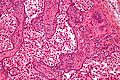
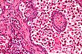
| LM | immature germ cells (resembling Sertoli cells or granulosa cells),[citation needed] primitive germ cells resemble those of a dysgerminoma, +/-calcification (very common), +/-eosinophilic basement membrane material between the (primitive) germ cells and support cells. |
| LM DDx | dysgerminoma |
| Site | ovary |
|
| |
Gonadoblastoma is a rare germ cell tumour with sex cord elements.
General
- Associated with abnormal sexual development.
- Often coexist with a dysgerminoma.
- A mixed tumour that consists of (1) primitive germ cells and (2) sex cord elements.
Gross
- +/-Cystic.
Microscopic
Features:[1]
- Immature germ cells resembling Sertoli cells or granulosa cells.[citation needed]
- Sertoli cells = moderate cytoplasm in a trabecular or tubular architecture.
- Granulosa cells = form follicle-like structures.
- May form nests.
- Primitive germ cells resemble those of a dysgerminoma.
- Polygonal cells with a central nucleus, squared-off nuclear membrane and clear cytoplasm.
- +/-Calcification (very common).
- +/-Eosinophilic basement membrane material between the (primitive) germ cells and support cells.[2]
Images
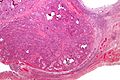
Gonadoblastoma - very low mag. (WC/Nephron)
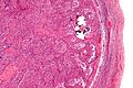
Gonadoblastoma - low mag. (WC/Nephron)
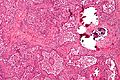
Gonadoblastoma - intermed mag. (WC/Nephron)
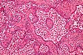
Gonadoblastoma - high mag. (WC/Nephron)
Gonadoblastoma - high mag. (WC/Nephron)
Gonadoblastoma - very high mag. (WC/Nephron)


www:
- Gonadoblastoma - low mag. (webpathology.com).
- Gonadoblastoma - high mag. (webpathology.com).
- Gonadoblastoma - high mag. (webpathology.com).
- Gonadoblastoma - low mag. (flickr.com).
- Gonadoblastoma - intermed. mag. (flickr.com).
- Gonadoblastoma - high mag. (flickr.com).
- Gonadoblastoma - several cases (upmc.edu).
See also
References
- ↑ Cotran, Ramzi S.; Kumar, Vinay; Fausto, Nelson; Nelso Fausto; Robbins, Stanley L.; Abbas, Abul K. (2005). Robbins and Cotran pathologic basis of disease (7th ed.). St. Louis, Mo: Elsevier Saunders. pp. 1104. ISBN 0-7216-0187-1.
- ↑ URL: http://www.flickr.com/photos/ckrishnan/3972432044/in/photostream/. Accessed on: 11 September 2011.