Difference between revisions of "Giant cell tumour of tendon sheath"
(redirect) |
|||
| (10 intermediate revisions by the same user not shown) | |||
| Line 1: | Line 1: | ||
{{ Infobox diagnosis | |||
| Name = {{PAGENAME}} | |||
| Image = Giant cell tumour of the tendon sheath -- very high mag.jpg | |||
| Width = | |||
| Caption = GCT of tendon sheath. [[H&E stain]]. | |||
| Micro = foam cells, multinucleated giant cells (may be scarce), +/-tendon, +/-hemosiderin-laden macrophages | |||
| Subtypes = | |||
| LMDDx = [[giant cell lesions]] | |||
| Stains = | |||
| IHC = | |||
| EM = | |||
| Molecular = | |||
| IF = | |||
| Gross = circumscribed mass - yellow-brown to tan | |||
| Grossing = | |||
| Site = hand - classic site | |||
| Assdx = | |||
| Syndromes = | |||
| Clinicalhx = | |||
| Signs = | |||
| Symptoms = | |||
| Prevalence = | |||
| Bloodwork = | |||
| Rads = | |||
| Endoscopy = | |||
| Prognosis = good (benign), can be malignant (rare) | |||
| Other = | |||
| ClinDDx = | |||
}} | |||
'''Giant cell tumour of tendon sheath''' is a relatively common tumour of small [[joints]]. It is grouped with the [[chondro-osseous tumours]]. It is abbreviated '''GCT of tendon sheath'''. | |||
''Fibroma of tendon sheath'' (abbreviated ''FTS'') redirect to this article. | |||
==General== | |||
*Can be thought of as the small joint version of [[diffuse tenosynovial giant-cell tumour]] ([[AKA]] ''PVNS'').<ref name=Ref_DCHH341>{{Ref DCHH|341-2}}</ref> | |||
*Rarely recur. | |||
*Classically afflicts the hand.<ref name=Ref_WMSP612>{{Ref WMSP|612}}</ref> | |||
*Rarely malignant.<ref name=pmid19094761>{{Cite journal | last1 = Pan | first1 = YW. | last2 = Huang | first2 = XY. | last3 = You | first3 = JF. | last4 = Tian | first4 = GL. | last5 = Li | first5 = C. | title = [Malignant giant cell tumor of the tendon sheaths in the hand]. | journal = Zhonghua Wai Ke Za Zhi | volume = 46 | issue = 21 | pages = 1645-8 | month = Nov | year = 2008 | doi = | PMID = 19094761 }}</ref><ref name=pmid8230758>{{Cite journal | last1 = Shinjo | first1 = K. | last2 = Miyake | first2 = N. | last3 = Takahashi | first3 = Y. | title = Malignant giant cell tumor of the tendon sheath: an autopsy report and review of the literature. | journal = Jpn J Clin Oncol | volume = 23 | issue = 5 | pages = 317-24 | month = Oct | year = 1993 | doi = | PMID = 8230758 }}</ref> | |||
==Gross== | |||
Features:<ref name=Ref_WMSP612>{{Ref WMSP|612}}</ref> | |||
*Circumscribed mass - yellow-brown to tan. | |||
Note: | |||
*May be associated with bony erosions in larger lesions.<ref name=Ref_WMSP612>{{Ref WMSP|612}}</ref> | |||
Image: | |||
*[http://www.ncbi.nlm.nih.gov/pmc/articles/PMC3122708/figure/Fig2/ GCT of tendon sheath (nih.gov)].<ref name=pmid22282671>{{Cite journal | last1 = Suresh | first1 = SS. | last2 = Zaki | first2 = H. | title = Giant cell tumor of tendon sheath: case series and review of literature. | journal = J Hand Microsurg | volume = 2 | issue = 2 | pages = 67-71 | month = Dec | year = 2010 | doi = 10.1007/s12593-010-0020-9 | PMID = 22282671 }}</ref> | |||
==Microscopic== | |||
Features:<ref name=Ref_DCHH341>{{Ref DCHH|341-2}}</ref> | |||
*Foam cells. | |||
**Cells with moderate to abundant foamy-appearing cytoplasm. | |||
*Multinucleated giant cells - may be scarce. | |||
*+/-Tendon. | |||
**Dense connective tissue. | |||
*+/-Hemosiderin-laden macrophages. | |||
Note: | |||
*Features of malignancy: nuclear pleomorphism,<ref name=pmid8230758>{{Cite journal | last1 = Shinjo | first1 = K. | last2 = Miyake | first2 = N. | last3 = Takahashi | first3 = Y. | title = Malignant giant cell tumor of the tendon sheath: an autopsy report and review of the literature. | journal = Jpn J Clin Oncol | volume = 23 | issue = 5 | pages = 317-24 | month = Oct | year = 1993 | doi = | PMID = 8230758 }}</ref> abnormal mitoses, >10 mitoses/[[HPF]], tumour necrosis lack of maturation to superficial part (nuclei shrink, cytoplasm lipid-ified).<ref name=Ref_DCHH341>{{Ref DCHH|341-2}}</ref> | |||
DDx: | |||
*[[Giant cell lesions]]. | |||
*Plexiform fibrohistiocytoma.{{fact}} | |||
*Fibroma of tendon sheath (FTS) - if one believes it is a separate entity.<ref>{{Cite journal | last1 = Heckert | first1 = R. | last2 = Bear | first2 = J. | last3 = Summers | first3 = T. | last4 = Frew | first4 = M. | last5 = Gwinn | first5 = D. | last6 = McKay | first6 = P. | title = Fibroma of the tendon sheath - a rare hand tumor. | journal = Pol Przegl Chir | volume = 84 | issue = 12 | pages = 651-6 | month = Dec | year = 2012 | doi = 10.2478/v10035-012-0107-z | PMID = 23399633 }}</ref> | |||
**IHC suggests ''FTS'' and ''GCT of tendon sheath'' are one entity.<Ref name=pmid7777476>{{Cite journal | last1 = Maluf | first1 = HM. | last2 = DeYoung | first2 = BR. | last3 = Swanson | first3 = PE. | last4 = Wick | first4 = MR. | title = Fibroma and giant cell tumor of tendon sheath: a comparative histological and immunohistological study. | journal = Mod Pathol | volume = 8 | issue = 2 | pages = 155-9 | month = Feb | year = 1995 | doi = | PMID = 7777476 }}</ref> | |||
===Images=== | |||
<gallery> | |||
Image: Giant cell tumour of the tendon sheath -- intermed mag.jpg | GCT of tendon sheath - intermed. mag. (WC) | |||
Image: Giant cell tumour of the tendon sheath -- high mag.jpg | GCT of tendon sheath - high mag. (WC) | |||
Image: Giant cell tumour of the tendon sheath -- very high mag.jpg | GCT of tendon sheath - very high mag. (WC) | |||
</gallery> | |||
<gallery> | |||
Image:Giant_cell_tumor_of_tendon_sheath_histopathology%281%29.jpg | GCT of tendon sheath. (WC/KGH) | |||
</gallery> | |||
www: | |||
*[http://www.webpathology.com/image.asp?n=1&Case=484 GCT of tendon sheath - very low mag. (webpathology.com)] | |||
*[http://www.webpathology.com/image.asp?case=484&n=5 GCT of tendon sheath - low mag. (webpathology.com)]. | |||
*[http://www.webpathology.com/image.asp?n=4&Case=484 GCT of tendon sheath - high mag. (webpathology.com)]. | |||
==Sign out== | |||
<pre> | |||
Submitted as "Giant Cell Tumour Right Long Finger", Excision: | |||
- Giant cell tumour of tendon sheath. | |||
</pre> | |||
===Block letters=== | |||
<pre> | |||
LESION, RIGHT INDEX FINGER, EXCISION: | |||
- GIANT CELL TUMOUR OF THE TENDON SHEATH. | |||
</pre> | |||
===Micro=== | |||
The sections show histiocytes and rare multinucleated giant cells on a background of dense connective tissue compatible with tendon. No nuclear atypia is apparent. Rare mitotic activity is identified. No atypical mitoses are apparent. | |||
====Alternate==== | |||
The sections show histiocyte-like cells and rare multinucleated giant cells on a background of dense | |||
connective tissue compatible with tendon. Hemosiderin-laden macrophages are present. No | |||
nuclear atypia is apparent. No mitotic activity is apparent. | |||
==See also== | |||
*[[Chondro-osseous tumours]]. | |||
*[[Diffuse tenosynovial giant-cell tumour]]. | |||
==References== | |||
{{Reflist|2}} | |||
[[Category:Diagnosis]] | [[Category:Diagnosis]] | ||
[[Category:Chondro-osseous tumours]] | |||
Latest revision as of 21:46, 11 October 2017
| Giant cell tumour of tendon sheath | |
|---|---|
| Diagnosis in short | |
 GCT of tendon sheath. H&E stain. | |
|
| |
| LM | foam cells, multinucleated giant cells (may be scarce), +/-tendon, +/-hemosiderin-laden macrophages |
| LM DDx | giant cell lesions |
| Gross | circumscribed mass - yellow-brown to tan |
| Site | hand - classic site |
|
| |
| Prognosis | good (benign), can be malignant (rare) |
Giant cell tumour of tendon sheath is a relatively common tumour of small joints. It is grouped with the chondro-osseous tumours. It is abbreviated GCT of tendon sheath.
Fibroma of tendon sheath (abbreviated FTS) redirect to this article.
General
- Can be thought of as the small joint version of diffuse tenosynovial giant-cell tumour (AKA PVNS).[1]
- Rarely recur.
- Classically afflicts the hand.[2]
- Rarely malignant.[3][4]
Gross
Features:[2]
- Circumscribed mass - yellow-brown to tan.
Note:
- May be associated with bony erosions in larger lesions.[2]
Image:
Microscopic
Features:[1]
- Foam cells.
- Cells with moderate to abundant foamy-appearing cytoplasm.
- Multinucleated giant cells - may be scarce.
- +/-Tendon.
- Dense connective tissue.
- +/-Hemosiderin-laden macrophages.
Note:
- Features of malignancy: nuclear pleomorphism,[4] abnormal mitoses, >10 mitoses/HPF, tumour necrosis lack of maturation to superficial part (nuclei shrink, cytoplasm lipid-ified).[1]
DDx:
- Giant cell lesions.
- Plexiform fibrohistiocytoma.[citation needed]
- Fibroma of tendon sheath (FTS) - if one believes it is a separate entity.[6]
- IHC suggests FTS and GCT of tendon sheath are one entity.[7]
Images
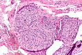
GCT of tendon sheath - intermed. mag. (WC)
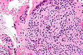
GCT of tendon sheath - high mag. (WC)
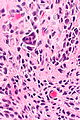
GCT of tendon sheath - very high mag. (WC)


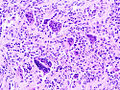
GCT of tendon sheath. (WC/KGH)
.jpg)
www:
- GCT of tendon sheath - very low mag. (webpathology.com)
- GCT of tendon sheath - low mag. (webpathology.com).
- GCT of tendon sheath - high mag. (webpathology.com).
Sign out
Submitted as "Giant Cell Tumour Right Long Finger", Excision:
- Giant cell tumour of tendon sheath.
Block letters
LESION, RIGHT INDEX FINGER, EXCISION:
- GIANT CELL TUMOUR OF THE TENDON SHEATH.
Micro
The sections show histiocytes and rare multinucleated giant cells on a background of dense connective tissue compatible with tendon. No nuclear atypia is apparent. Rare mitotic activity is identified. No atypical mitoses are apparent.
Alternate
The sections show histiocyte-like cells and rare multinucleated giant cells on a background of dense connective tissue compatible with tendon. Hemosiderin-laden macrophages are present. No nuclear atypia is apparent. No mitotic activity is apparent.
See also
References
- ↑ 1.0 1.1 1.2 Tadrous, Paul.J. Diagnostic Criteria Handbook in Histopathology: A Surgical Pathology Vade Mecum (1st ed.). Wiley. pp. 341-2. ISBN 978-0470519035.
- ↑ 2.0 2.1 2.2 Humphrey, Peter A; Dehner, Louis P; Pfeifer, John D (2008). The Washington Manual of Surgical Pathology (1st ed.). Lippincott Williams & Wilkins. pp. 612. ISBN 978-0781765275.
- ↑ Pan, YW.; Huang, XY.; You, JF.; Tian, GL.; Li, C. (Nov 2008). "[Malignant giant cell tumor of the tendon sheaths in the hand].". Zhonghua Wai Ke Za Zhi 46 (21): 1645-8. PMID 19094761.
- ↑ 4.0 4.1 Shinjo, K.; Miyake, N.; Takahashi, Y. (Oct 1993). "Malignant giant cell tumor of the tendon sheath: an autopsy report and review of the literature.". Jpn J Clin Oncol 23 (5): 317-24. PMID 8230758.
- ↑ Suresh, SS.; Zaki, H. (Dec 2010). "Giant cell tumor of tendon sheath: case series and review of literature.". J Hand Microsurg 2 (2): 67-71. doi:10.1007/s12593-010-0020-9. PMID 22282671.
- ↑ Heckert, R.; Bear, J.; Summers, T.; Frew, M.; Gwinn, D.; McKay, P. (Dec 2012). "Fibroma of the tendon sheath - a rare hand tumor.". Pol Przegl Chir 84 (12): 651-6. doi:10.2478/v10035-012-0107-z. PMID 23399633.
- ↑ Maluf, HM.; DeYoung, BR.; Swanson, PE.; Wick, MR. (Feb 1995). "Fibroma and giant cell tumor of tendon sheath: a comparative histological and immunohistological study.". Mod Pathol 8 (2): 155-9. PMID 7777476.