Difference between revisions of "Gaucher disease"
Jump to navigation
Jump to search
(→Microscopic: +images) |
(→Images: +image) |
||
| Line 59: | Line 59: | ||
===Images=== | ===Images=== | ||
<gallery> | <gallery> | ||
Image:Gaucher disease - intermed mag.jpg | Gaucher disease - intermed. mag. (WC) | |||
Image:Gaucher_disease_-_high_mag.jpg | Gaucher disease - high mag. (WC) | |||
Image:Gaucher_disease_-_very_high_mag.jpg | Gaucher disease - with fine vesicular cytoplasm - very high mag. (WC) | Image:Gaucher_disease_-_very_high_mag.jpg | Gaucher disease - with fine vesicular cytoplasm - very high mag. (WC) | ||
</gallery> | </gallery> | ||
www: | www: | ||
Revision as of 09:05, 29 July 2013
| Gaucher disease | |
|---|---|
| Diagnosis in short | |
 Gaucher disease. H&E stain. | |
|
| |
| LM | crumpled tissue paper macrophages |
| Subtypes | type I, type II, type III |
| Site | bone, other |
|
| |
| Associated Dx | fracture of bone |
| Blood work | pancytopenia |
| Prognosis | dependent on subtype |
Gaucher disease a lysosomal storage disease. It is a rare thingy that may be seen in people that marry their cousins.
Pathology
- Accumulation of glucocerebroside in monocytes/macrophages due to deficiency of glucocerebrosidase.[1]
- Defect in acid beta-glucosidase gene (GBA gene).[2][3][4]
Subtypes
- There are several types - all are autosomal recessive.[1]
Types:[5]
- Type I: 99% of cases; no CNS involvement - survive to adulthood.
- Type II: infantile onset - CNS degeneration + death at young age.
- Type III: mixed of type I & type II.
Clinical
- Pancytopenia - due to marrow replacement.
- Hepatosplenomegaly (type I).
Microscopic
- Mononuclear phagocytes with abundant eosinophilic cytoplasm with subtle irregular lines (~0.5 micrometers in width).
- Known as "crumpled tissue paper cells" / "crumpled tissue paper cytoplasm."[7]
Notes:
- Crumpled tissue paper: crumpled tissue paper - image (123rf.com).
- The textbook case may look crumpled... along with some mind altering drugs.
- The typical case is:
- Abundant macrophages with cytoplasm filled by very small (clear) vacuoles (~0.2-0.4 micrometres).
- The typical case is:
Images
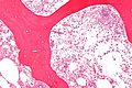
Gaucher disease - intermed. mag. (WC)
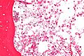
Gaucher disease - high mag. (WC)
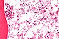
Gaucher disease - with fine vesicular cytoplasm - very high mag. (WC)


www:
- Gaucher disease - bone marrow aspirate (swmed.edu).
- Gaucher disease (webpathology.com).[6]
- Gaucher disease (neuropathologyweb.org).[8]
Stains
- Material in "crumpled tissue paper cells": PAS +ve.[5]
See also
References
- ↑ 1.0 1.1 URL: http://emedicine.medscape.com/article/944157-overview. Accessed on: 3 December 2010.
- ↑ Online 'Mendelian Inheritance in Man' (OMIM) 230800
- ↑ Online 'Mendelian Inheritance in Man' (OMIM) 230900
- ↑ Online 'Mendelian Inheritance in Man' (OMIM) 231000
- ↑ 5.0 5.1 5.2 Mitchell, Richard; Kumar, Vinay; Fausto, Nelson; Abbas, Abul K.; Aster, Jon (2011). Pocket Companion to Robbins & Cotran Pathologic Basis of Disease (8th ed.). Elsevier Saunders. pp. 95. ISBN 978-1416054542.
- ↑ 6.0 6.1 URL: http://www.webpathology.com/image.asp?case=377&n=3. Accessed on: 30 November 2010.
- ↑ URL: http://pathcuric1.swmed.edu/pathdemo/gen1/gen130.htm. Accessed on: 28 May 2011.
- ↑ URL: http://www.neuropathologyweb.org/chapter10/chapter10bLSDs.html. Accessed on: 30 November 2010.