Flat epithelial atypia
Jump to navigation
Jump to search
The printable version is no longer supported and may have rendering errors. Please update your browser bookmarks and please use the default browser print function instead.
Flat epithelial atypia, abbreviated FEA, is a benign breast lesion that is associated with an increased risk of subsequent breast cancer.
General
Epidemiology:
- Associated with ADH & DCIS; may represent a non-obligate precursor lesion of ADH & DCIS.[1]
- Low risk of progression to invasive malignancy.[2]
Management:
- Excision.
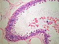
Microscopic
Features:
- "Flat" ~ three cells thick.
- Hypercellular gland -- several layers.
- Columnar cell morphology.
- +/-Apical snouts.
DDx:
- Columnar cell change.
- Columnar cell hyperplasia.
- ADH.
- Flat DCIS (clinging carcinoma).
- Apocrine cyst - granular cytoplasm.
- Tubular carcinoma - should be considered due to the association.
Images
Breast - Flat Atypia (SKB)
_PA.JPG)
Molecular
- Loss of 16q.
- Not used for diagnosis.
See also
References
- ↑ Lerwill, MF. (Apr 2008). "Flat epithelial atypia of the breast.". Arch Pathol Lab Med 132 (4): 615-21. doi:10.1043/1543-2165(2008)132[615:FEAOTB]2.0.CO;2. PMID 18384213.
- ↑ Schnitt, SJ. (2003). "The diagnosis and management of pre-invasive breast disease: flat epithelial atypia--classification, pathologic features and clinical significance.". Breast Cancer Res 5 (5): 263-8. doi:10.1186/bcr625. PMID 12927037.