Epidermal inclusion cyst
| Epidermal inclusion cyst | |
|---|---|
| Diagnosis in short | |
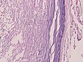
 Epidermal inclusion cyst. H&E stain. | |
|
| |
| LM | cyst lining by a stratified squamous epithelium with a granular layer - contains keratin; no significant nuclear atypia; +/-granulomatous inflammation (due to rupture) |
| LM DDx | pilar cyst, eccrine hidrocystoma (eyelid), dermoid cyst, cystic squamous cell carcinoma, keratoacanthoma, dermatofibrosarcoma protuberans, hybrid epidermoid and trichilemmal cyst |
| Gross | nodule +/-yellow colour |
| Site | skin |
|
| |
| Prevalence | very common |
| Prognosis | benign |
| Clin. DDx | other skin cysts, gouty tophus |
Epidermal inclusion cyst, abbreviated EIC, is a very common skin pathology. It is also know as epidermal cyst, epidermoid cyst,[1] and follicular cyst, infundibular type.
Testicular epidermoid cyst is dealt with separately in epidermoid cyst of the testis.
General
- Very common.
- The clinical term is sebaceous cyst.
- This is a misnomer as they contain keratin (not sebum).[2][3]
- The term may be used to refer to a pilar cyst.
Gross
Features:[4]
- Nodule.
- +/-Yellowish colour.
DDx:
- Pilar cyst - indistinguishable on gross.[2]
- Melanoma.[3]
- Gouty tophus.
Image
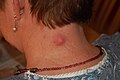
Epidermal inclusion cyst. (WC)

Microscopic
Features:
- Cyst lining by a stratified squamous epithelium with a granular layer - key feature.[5]
- No significant nuclear atypia.
- Contains keratin - acellular, lamellar appearance.
- +/-Granulomatous inflammation due to rupture.
DDx:
- Pilar cyst - no granular layer.
- Eccrine hidrocystoma - eyelid lesion; same histology.[6]
- Dermoid cyst - has adnexal structures, i.e. hair follicle, sebaceous glands, sweat glands.
- Cystic squamous cell carcinoma.[7]
- Keratoacanthoma.[1]
- Dermatofibrosarcoma protuberans - if lesion is large.
- Hybrid epidermoid and trichilemmal cyst.[8]
Images
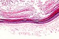
EIC - intermed. mag. (WC)
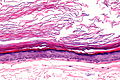
EIC - high mag. (WC)
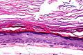
EIC - very high mag. (WC)


Dermoid cyst - intracranial. (WC)

www:
- Epidermal inclusion cyst (flickr.com).
- Epidermal inclusion cyst (ajronline.org).[9]
- Ruptured epidermal inclusion cyst (nih.gov).
Sign out
SKIN CYST, BACK, EXCISION:
- EPIDERMAL INCLUSION CYST.
Ruptured
Lesion, Left Elbow, Excision:
- Benign epidermal inclusion cyst with evidence of rupture.
Block letters
SKIN LESION, RIGHT CHEEK, EXCISION:
- RUPTURED EPIDERMAL INCLUSION CYST.
Overlying skin
SKIN LESION (QUERY CYST), RIGHT JAW, EXCISION:
- EPIDERMAL INCLUSION CYST.
- BENIGN (OVERLYING) SKIN WITH SOLAR ELASTOSIS.
- NEGATIVE FOR DYSPLASIA AND NEGATIVE FOR MALIGNANCY.
Not quite a cyst
"CYST", LEFT INDEX FINGER, EXCISION:
- LARGE (CYST-LIKE) EPIDERMAL INVAGINATION CONTAINING KERATIN.
- DERMAL SCAR.
- MINIMAL (NONSPECIFIC) PERIVASCULAR INFLAMMATION.
- NO DEFINITE CYST APPARENT.
- NEGATIVE FOR DYSPLASIA AND NEGATIVE FOR MALIGNANCY.
Micro
The sections show hair-bearing skin with a cyst that is lined by squamous epithelium with a granular layer. The cyst contains keratin. The overlying epithelium is unremarkable.
Ruptured
The sections show hair-bearing skin with a cyst that is lined by squamous epithelium with a granular layer. The cyst contains keratin. A mixed inflammatory infiltrate (predominantly lymphocytes and plasma cells) surround the cyst. Neutrophils infiltrate the cyst lining and are admixed with the keratin within its core.
The lesion appears to be completely excised in the plane of section. Hair follicles are adjacent to the lesion; however, they are not inflamed. The overlying epithelium is unremarkable.
Ruptured without epithelium
The section shows a dermal collection of neutrophils with acellular keratin-like material surrounded by histiocytes and fibrosis. The lesion is completely excised in the plane of section. Hair follicles are adjacent to the abscess; however, they are not inflamed.
See also
References
- ↑ 1.0 1.1 Busam, Klaus J. (2009). Dermatopathology: A Volume in the Foundations in Diagnostic Pathology Series (1st ed.). Saunders. pp. 302. ISBN 978-0443066542.
- ↑ 2.0 2.1 URL: http://www.dermis.net/dermisroot/en/36946/diagnose.htm. Accessed on: 2 November 2012.
- ↑ 3.0 3.1 Venus, MR.; Eltigani, EA.; Fagan, JM. (Sep 2007). "Just another sebaceous cyst?". Ann R Coll Surg Engl 89 (6): W19-21. doi:10.1308/147870807X227791. PMC 2121251. PMID 18201468. https://www.ncbi.nlm.nih.gov/pmc/articles/PMC2121251/..
- ↑ URL: http://dermatlas.med.jhmi.edu/derm/result.cfm?diagnosis=128. Accessed on: 2 November 2012.
- ↑ URL: http://emedicine.medscape.com/article/1058907-diagnosis. Accessed on: 18 March 2011.
- ↑ Adams, SP. (Feb 1999). "Dermacase. Eccrine hydrocystoma.". Can Fam Physician 45: 297, 306. PMC 2328272. PMID 10065300. https://www.ncbi.nlm.nih.gov/pmc/articles/PMC2328272/.
- ↑ Lin, CY.; Jwo, SC. (Apr 2002). "Squamous cell carcinoma arising in an epidermal inclusion cyst.". Chang Gung Med J 25 (4): 279-82. PMID 12079164.
- ↑ Brownstein, MH. (Dec 1983). "Hybrid cyst: a combined epidermoid and trichilemmal cyst.". J Am Acad Dermatol 9 (6): 872-5. PMID 6643785.
- ↑ Crystal, P.; Shaco-Levy, R. (Mar 2005). "Concentric rings within a breast mass on sonography: lamellated keratin in an epidermal inclusion cyst.". AJR Am J Roentgenol 184 (3 Suppl): S47-8. PMID 15728019.