Emphysema
Jump to navigation
Jump to search
The printable version is no longer supported and may have rendering errors. Please update your browser bookmarks and please use the default browser print function instead.
| Emphysema | |
|---|---|
| Diagnosis in short | |
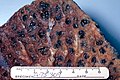
 Emphysematous changes. H&E stain. | |
|
| |
| LM | alveoli too large, thin septa (no interstitial thickening) |
| Subtypes | centriacinar (centrilobular) emphysema, panacinar (panlobular) emphysema, distal (paraseptal) acinar emphysema, irregular emphysema |
| Gross | usually upper lobe predominant - blebs, bullae |
| Site | lung |
|
| |
| Associated Dx | +/-pneumothorax |
| Syndromes | Alpha-1 antitrypsin deficiency, others |
|
| |
| Clinical history | +/-smoking |
| Signs | barrel-shaped chest |
| Symptoms | shortness of breath |
| Prevalence | common |
| Radiology | hyperinflation, Saber-sheath trachea (associated with COPD) |
| Prognosis | dependent on underlying cause |
| Treatment | stop smoking, bullectomy |
Emphysema is a common medical lung disease strongly associated with smoking.
Chronic obstructive pulmonary disease, abbreviated COPD, redirects here.
General
- Usually due to smoking.
- Often lumped together with chronic bronchitis and called chronic obstructive pulmonary disease (COPD).[1]
- May cause pneumothorax - especially in young adults.[2]
Causes of emphysema other than smoking:[3]
Pathologic classification
Based on morphology:[4]
- Centriacinar (centrilobular) emphysema - associated with heavy smoking.
- Panacinar (panlobular) emphysema - associated with alpha-1 antitrypsin deficiency.
- Distal (paraseptal) acinar emphysema - associated with spontaneous pneumothorax.
- Irregular emphysema - usu. insignificant.
Note:
- Why does smoking lead to centriacinar emphysema?
- The bad stuff from smoking gets enters the acinus at the centre; ergo, this is the location of the most damage.
Gross
- Holes (blebs, bullae), usually upper lung field predominant.
- Lungs may overlap the heart.[5]
Notes:
Images
Centrilobular emphysema. (WC/Edwin Ewing Jr.)

Radiology
- Saber-sheath trachea - a finding associated with COPD.[8]
- Trachea's anterior to posterior dimension:left to right dimension is >2:1.[9]
- Barrel-shaped chest.[10]
Microscopic
Features:[5]
- Large alveoli.
- Thin septa (no interstitial thickening).
Images
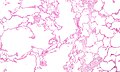
Emphysema. (WC)
See also
References
- ↑ Mitchell, Richard; Kumar, Vinay; Fausto, Nelson; Abbas, Abul K.; Aster, Jon (2011). Pocket Companion to Robbins & Cotran Pathologic Basis of Disease (8th ed.). Elsevier Saunders. pp. 368. ISBN 978-1416054542.
- ↑ Leslie, Kevin O.; Wick, Mark R. (2004). Practical Pulmonary Pathology: A Diagnostic Approach (1st ed.). Churchill Livingstone. pp. 296. ISBN 978-0443066313.
- ↑ Lee, P.; Gildea, TR.; Stoller, JK. (Dec 2002). "Emphysema in nonsmokers: alpha 1-antitrypsin deficiency and other causes.". Cleve Clin J Med 69 (12): 928-9, 933, 936 passim. PMID 12546267.
- ↑ Mitchell, Richard; Kumar, Vinay; Fausto, Nelson; Abbas, Abul K.; Aster, Jon (2011). Pocket Companion to Robbins & Cotran Pathologic Basis of Disease (8th ed.). Elsevier Saunders. pp. 368. ISBN 978-1416054542.
- ↑ 5.0 5.1 Mitchell, Richard; Kumar, Vinay; Fausto, Nelson; Abbas, Abul K.; Aster, Jon (2011). Pocket Companion to Robbins & Cotran Pathologic Basis of Disease (8th ed.). Elsevier Saunders. pp. 369. ISBN 978-1416054542.
- ↑ URL: http://dictionary.reference.com/browse/bleb. Accessed on: 3 August 2011.
- ↑ URL: http://dictionary.reference.com/browse/bulla. Accessed on: 3 August 2011.
- ↑ Tunsupon P, Dhillon SS, Harris K, Alraiyes AH (February 2016). "Saber-sheath trachea in a patient with severe COPD". BMJ Case Rep 2016. doi:10.1136/bcr-2016-214648. PMC 4769447. PMID 26912770. https://www.ncbi.nlm.nih.gov/pmc/articles/PMC4769447/.
- ↑ Webb EM, Elicker BM, Webb WR (May 2000). "Using CT to diagnose nonneoplastic tracheal abnormalities: appearance of the tracheal wall". AJR Am J Roentgenol 174 (5): 1315–21. doi:10.2214/ajr.174.5.1741315. PMID 10789785.
- ↑ "Physical signs in patients with chronic obstructive pulmonary disease". Lung India 36 (1): 38–47. 2019. doi:10.4103/lungindia.lungindia_145_18. PMC 6330798. PMID 30604704. https://www.ncbi.nlm.nih.gov/pmc/articles/PMC6330798/.