Embryonal carcinoma
Jump to navigation
Jump to search
The printable version is no longer supported and may have rendering errors. Please update your browser bookmarks and please use the default browser print function instead.
| Embryonal carcinoma | |
|---|---|
| Diagnosis in short | |
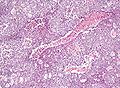
 Embryonal carcinoma. H&E stain. | |
|
| |
| LM | vesicular nuclei, nuclear overlap, necrosis (common), mitoses, variable architecture (tubulopapillary, glandular, solid, embryoid bodies) |
| LM DDx | seminoma, mixed germ cell tumour, yolk sac tumour, other carcinomas |
| IHC | OCT3 +ve, CD30 +ve, AE1/AE3 +ve, CD117 -ve |
| Grossing notes | orchiectomy grossing |
| Staging | testicular cancer staging |
| Site | testis, ovary, mediastinum |
|
| |
| Signs | testicular mass, pelvic mass |
| Prevalence | pure embryonal uncommon |
| Clin. DDx | typically other testicular tumours |
Embryonal carcinoma is a type of germ cell tumour. It is commonly as a component of mixed germ cell tumours.
General
- Affects young adults.
- May be seen in women.
- Usually a component of a mixed germ cell tumour - in the testicle 85% of cases are mixed, only 15% are pure.[1]
Gross
- Typically a testicular mass.
- May be seen in the mediastinum.[2]
Microscopic
Features:[3]
- Nucleoli - key feature.
- Vesicular nuclei (clear, empty appearing nuclei) - key feature.
- Nuclei overlap.
- Necrosis - common.
- Not commonly present in seminoma.
- Indistinct cell borders
- Mitoses - common.
- Variable architecture:[1]
- Solid (predominant in ~55% of cases).
- Glandular (predominant in ~17% of cases).
- Papillary (predominant in ~11% of cases).
- Nested.
- Micropapillary.
- Anastomosing glandular.
- Sieve-like glandular.
- Pseudopapillary.
- Blastocyst-like.
- Embryoid bodies - ball of cells in surrounded by empty space on three sides.
Notes:
- Cytoplasmic staining variable (eosinophilic to basophilic).
- Syncytiotrophoblasts commonly seen (~40-50% of cases[1]).
DDx:
Images
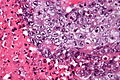
Embryonal carcinoma - very high mag. - cropped (WC/Nephron)
Embryonal carcinoma - high mag. (WC/Nephron)

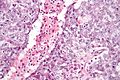
Embryonal carcinoma - high mag. (WC/Nephron)
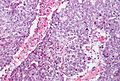
Embryonal carcinoma - intermed. mag. (WC/Nephron)
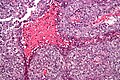
Embryonal carcinoma - low mag. (WC/Nephron)


IHC
ISUP consensus
General panel:[4]
- OCT4 +ve.
- Choriocarcinoma, yolk sac tumour and spermatocytic seminoma all -ve.
- CD30 +ve.
- -ve in seminoma.
- CD117 -ve.[5]
- +ve in seminoma.
Additional notes
- CK19 -ve.[6]
- Yolk sac tumour +ve, seminoma -ve, GCNIS (ITGCN) -ve, normal testis -ve.
- AE1/AE3 +ve.
- OCT3/4 +ve.[7]
- D2-40 -ve.[7]
- PLAP +ve.[8]
See also
References
- ↑ 1.0 1.1 1.2 Kao, CS.; Ulbright, TM.; Young, RH.; Idrees, MT. (May 2014). "Testicular embryonal carcinoma: a morphologic study of 180 cases highlighting unusual and unemphasized aspects.". Am J Surg Pathol 38 (5): 689-97. doi:10.1097/PAS.0000000000000171. PMID 24503753.
- ↑ Yalçın, B.; Demir, HA.; Tanyel, FC.; Akçören, Z.; Varan, A.; Akyüz, C.; Kutluk, T.; Büyükpamukçu, M. (Oct 2012). "Mediastinal germ cell tumors in childhood.". Pediatr Hematol Oncol 29 (7): 633-42. doi:10.3109/08880018.2012.713084. PMID 22877235.
- ↑ Zhou, Ming; Magi-Galluzzi, Cristina (2006). Genitourinary Pathology: A Volume in Foundations in Diagnostic Pathology Series (1st ed.). Churchill Livingstone. pp. 549. ISBN 978-0443066771.
- ↑ Ulbright TM, Tickoo SK, Berney DM, Srigley JR (August 2014). "Best practices recommendations in the application of immunohistochemistry in testicular tumors: report from the international society of urological pathology consensus conference". Am. J. Surg. Pathol. 38 (8): e50–9. doi:10.1097/PAS.0000000000000233. PMID 24832161.
- ↑ 5.0 5.1 Lau, SK.; Weiss, LM.; Chu, PG. (Mar 2007). "D2-40 immunohistochemistry in the differential diagnosis of seminoma and embryonal carcinoma: a comparative immunohistochemical study with KIT (CD117) and CD30.". Mod Pathol 20 (3): 320-5. doi:10.1038/modpathol.3800749. PMID 17277761.
- ↑ Bremmer, F.; Ströbel, P.; Hubertus, H.; Strecker, J.; Gaisa, N.; Strauß, A.; Schweyer, S.; Radzun, H-J. et al. (2015). [www.diagnosticpathology.org/content/10/1/7Cached "CK19 is a sensitive marker for yolk sac tumours of the testis."]. Diagnostic Pathology 10 (7). doi:10.1186/s13000-015-0243-y. www.diagnosticpathology.org/content/10/1/7Cached.
- ↑ 7.0 7.1 Iczkowski, KA.; Butler, SL.; Shanks, JH.; Hossain, D.; Schall, A.; Meiers, I.; Zhou, M.; Torkko, KC. et al. (Feb 2008). "Trials of new germ cell immunohistochemical stains in 93 extragonadal and metastatic germ cell tumors.". Hum Pathol 39 (2): 275-81. doi:10.1016/j.humpath.2007.07.002. PMID 18045648.
- ↑ Jacobsen, GK.; Nørgaard-Pedersen, B. (Sep 1984). "Placental alkaline phosphatase in testicular germ cell tumours and in carcinoma-in-situ of the testis. An immunohistochemical study.". Acta Pathol Microbiol Immunol Scand A 92 (5): 323-9. PMID 6209917.