Dermatofibrosarcoma protuberans
Jump to navigation
Jump to search
The printable version is no longer supported and may have rendering errors. Please update your browser bookmarks and please use the default browser print function instead.
| Dermatofibrosarcoma protuberans | |
|---|---|
| Diagnosis in short | |
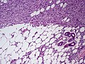
 DFSP. H&E stain. | |
|
| |
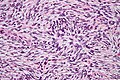
| LM | dermal spindle cell lesion with storiform pattern, typically contains adipose tissue within the tumour -- described as "honeycomb pattern" and "Swiss cheese pattern" |
| LM DDx | dermatofibroma, dermatomyofibroma, nodular fasciitis |
| IHC | CD34 +ve, Factor XIIIa -ve |
| Molecular | t(17;22)(q22;q15) |
| Gross | firm plaque +/-ulceration |
| Site | skin - usually trunk or proximal extremities |
|
| |
| Clinical history | second to fifth decade |
| Prevalence | uncommon |
| Prognosis | moderate, locally aggressive, rarely metastases |
| Treatment | wide excision |
Dermatofibrosarcoma protuberans, abbreviated DFSP, is a rare locally aggressive tumour of the skin.
General
- Destroys adnexal structures - somewhat unusual for a mostly benign tumour.
- Occasionally transforms to a (more aggressive) fibrosarcoma.[1]
- Typically slow growing.[2]
- Usually second to fifth decade.[2]
Treatment:[3]
- Wide excision.
- May include imatinib (Gleevec).
Gross
Features:[4]
- Firm plaque, often bosselated, usually on the trunk.
- +/-Ulceration.
Images:
- Protuberant DFSP (dermatlas.med.jhmi.edu).
- Huge DFSP on back (dermatlas.med.jhmi.edu).
- Protuberant DFSP - gross and histology (dermatlas.med.jhmi.edu).
Microscopic
Features:[3]
- Dermal spindle cell lesion with storiform pattern.
- Spokes of the wheel-pattern.
- Contains adipose tissue within the tumour -- key feature.
- Described as "honeycomb pattern" and "Swiss cheese pattern".
Notes:
- Adnexal structure within tumour are preserved -- this is unusual for a malignant tumour -- important.
DDx:
- Dermatofibroma - main DDx - has entrapment of collagen bundles at the edge of the lesion.
- Dermatomyofibroma.[5]
- Nodular fasciitis.
DDx of storiform pattern:
- DFSP.
- Dermatofibroma.
- Solitary fibrous tumour.
- Undifferentiated pleomorphic sarcoma.
Subtypes
Numerous variants/subtypes are described:[2]
- Pigmented DFSP (Bednar tumour).
- Myxoid DFSP.
- Myoid DFSP.
- Granular cell DFSP.
- Sclerotic DFSP.
- Atrophic DFSP,
- Giant cell fibroblastoma.
- DFSP with fibrosarcomatous areas.
Images
DFSP with fat entrapped. (WC)
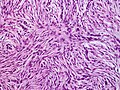
DFSP - high mag. (WC)
DFSP - storiform pattern - intermed. mag. (WC/Nephron)
DFSP - storiform pattern - very high mag. (WC/Nephron)


www:
IHC
Panel:[6]
- CD34 +ve.
- Factor XIIIa -ve.
- S-100 -ve (screen for melanoma).
- Caldesmin -ve (screen for muscle differentiation).
- Beta-catenin. (???)
- MIB1 (proliferation marker).
- Should not be confused with MIB-1 a gene that regulates apoptosis.
Molecular
A characteristic translocation is seen:[9] t(17;22)(q22;q15) COLA1/PDGFB.
See also
References
- ↑ Stacchiotti, S.; Pedeutour, F.; Negri, T.; Conca, E.; Marrari, A.; Palassini, E.; Collini, P.; Keslair, F. et al. (Oct 2011). "Dermatofibrosarcoma protuberans-derived fibrosarcoma: clinical history, biological profile and sensitivity to imatinib.". Int J Cancer 129 (7): 1761-72. doi:10.1002/ijc.25826. PMID 21128251.
- ↑ 2.0 2.1 2.2 Llombart, B.; Serra-Guillén, C.; Monteagudo, C.; López Guerrero, JA.; Sanmartín, O. (Feb 2013). "Dermatofibrosarcoma protuberans: a comprehensive review and update on diagnosis and management.". Semin Diagn Pathol 30 (1): 13-28. doi:10.1053/j.semdp.2012.01.002. PMID 23327727.
- ↑ 3.0 3.1 Kumar, Vinay; Abbas, Abul K.; Fausto, Nelson; Aster, Jon (2009). Robbins and Cotran pathologic basis of disease (8th ed.). Elsevier Saunders. pp. 1183. ISBN 978-1416031215.
- ↑ Mitchell, Richard; Kumar, Vinay; Fausto, Nelson; Abbas, Abul K.; Aster, Jon (2011). Pocket Companion to Robbins & Cotran Pathologic Basis of Disease (8th ed.). Elsevier Saunders. pp. 600. ISBN 978-1416054542.
- ↑ Busam, Klaus J. (2009). Dermatopathology: A Volume in the Foundations in Diagnostic Pathology Series (1st ed.). Saunders. pp. 504. ISBN 978-0443066542.
- ↑ AP. May 2009.
- ↑ 7.0 7.1 Abenoza P, Lillemoe T (October 1993). "CD34 and factor XIIIa in the differential diagnosis of dermatofibroma and dermatofibrosarcoma protuberans". Am J Dermatopathol 15 (5): 429–34. PMID 7694515.
- ↑ 8.0 8.1 Goldblum JR, Tuthill RJ (April 1997). "CD34 and factor-XIIIa immunoreactivity in dermatofibrosarcoma protuberans and dermatofibroma". Am J Dermatopathol 19 (2): 147–53. PMID 9129699.
- ↑ Kumar, Vinay; Abbas, Abul K.; Fausto, Nelson; Aster, Jon (2009). Robbins and Cotran pathologic basis of disease (8th ed.). Elsevier Saunders. pp. 1249. ISBN 978-1416031215.