Dermal scar
Jump to navigation
Jump to search
The printable version is no longer supported and may have rendering errors. Please update your browser bookmarks and please use the default browser print function instead.
| Dermal scar | |
|---|---|
| Diagnosis in short | |
 Dermal scar. H&E stain. | |
|
| |
| LM | dense collagen - fibers run parallel to the DE junction, loss of dermal papilla, loss of adnexal structures, thin-wall blood vessels |
| LM DDx | malignant melanoma desmoplastic-neurotropic type, dermatofibroma, desmoplastic Spitz nevus, sclerosing blue nevus |
| Stains | S-100 -ve (mostly) |
| Site | skin |
|
| |
| Clinical history | trauma, previous excision or biopsy |
| Prevalence | common |
| Prognosis | benign |
| Dermal scar | |
|---|---|
| External resources | |
| EHVSC | 10187 (Dermal scar adjacent to a basal cell carcinoma) |
| Wikipedia | Scar |
Dermal scar, also simply scar, is commonly seen in dermatopathology. It is also known a cicatrix.
General
- Previous surgery, biopsy, trauma.
Gross
Images
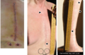
Scars. (WC)

Microscopic
Features:
- Loss of dermal papilla.
- Dense collagen - fibers run parallel to the dermal-epidermal (DE) junction[3] - key feature.
- Loss of adnexal structures.
Other feature:
- Thin-walled blood vessels.
- Described as running perpendicular to the surface[3] - this may not be apparent.
Note:
- There should not be any nuclear hyperchromasia or pleomorphism.[4]
DDx:
- Malignant melanoma, desmoplastic-neurotropic type - nuclear pleomorphism and/or hyperchromasia; may be focal.[4]
- Epidermal hyperplasia and the preservation of adnexal structures is very suspicious.
- Dermatofibroma.
- Desmoplastic Spitz nevus.
- Sclerosing blue nevus.
Image
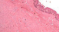
Scar. (WC)
IHC
- S100 focal/scattered +ve.
- Desmoplastic melanoma strong +ve.
- HMB-45 -ve.
- Sclerosing blue nevus +ve.
Sign out
SKIN, LOWER MID BACK, RE-EXCISION:
- DERMAL SCAR.
- SOLAR ELASTOSIS.
SKIN LESION, LEFT UPPER ABDOMINAL WALL, RE-EXCISION:
- DERMAL SCAR, COMPLETELY EXCISED.
- BENIGN PIGMENT.
- NEGATIVE FOR DYSPLASIA AND NEGATIVE FOR MALIGNANCY.
Micro
The sections show skin with a dermis with dense collagen fibres that run parallel to the skin surface without adnexal structures. The overlying dermal-epidermis interface lacks the typical undulation.
See also
References
- ↑ Velangi, SS.; Rees, JL.. "Why are scars pale? An immunohistochemical study indicating preservation of melanocyte number and function in surgical scars.". Acta Derm Venereol 81 (5): 326-8. PMID 11800137.
- ↑ Chadwick, S.; Heath, R.; Shah, M. (May 2012). "Abnormal pigmentation within cutaneous scars: A complication of wound healing.". Indian J Plast Surg 45 (2): 403-11. doi:10.4103/0970-0358.101328. PMID 23162241.
- ↑ 3.0 3.1 Busam, Klaus J. (2009). Dermatopathology: A Volume in the Foundations in Diagnostic Pathology Series (1st ed.). Saunders. pp. 499. ISBN 978-0443066542.
- ↑ 4.0 4.1 Busam, Klaus J. (2009). Dermatopathology: A Volume in the Foundations in Diagnostic Pathology Series (1st ed.). Saunders. pp. 479. ISBN 978-0443066542.