Cutaneous calcinosis
Jump to navigation
Jump to search
The printable version is no longer supported and may have rendering errors. Please update your browser bookmarks and please use the default browser print function instead.
| Cutaneous calcinosis | |
|---|---|
| Diagnosis in short | |
 Cutaneous calcinosis. H&E stain. | |
|
| |
| Synonyms | cutaneous calcification, calcinosis cutis |
|
| |
| LM | dermal calcification - usu. well-circumscribed |
| Gross | firm nodule |
| Site | skin, scrotum |
|
| |
| Clinical history | +/-trauma at the site |
| Signs | firm nodule |
| Prevalence | uncommon |
| Prognosis | benign |
| Treatment | excision |
Cutaneous calcinosis, also calcinosis cutis and cutaneous calcification, is calcification of the skin. It is benign in itself; however, the underlying cause may not be.
General
- Benign in itself; underlying cause may not be benign.
- May be a scrotal lesion - known as scrotal calcinosis.[1]
Subtypes:[2]
- Dystrophic - due to death of cells; may be related to a tumour.
- Metastatic - due to chronic renal failure; hyperkalemia; paraneoplastic phenomenon.
- Iatrogenic - post surgical.
- Idiopathic.
Gross
- Firm nodule.
Microscopic
Features:
- Dermal calcification:
- Acellular purple blobs on H&E.
- +/-Artefactual tearing of surrounding tissue due to processing (cutting).
- +/-Small artefactual lines ~1-2 micrometers due to processing (cutting).
- +/-Greyish rim of paucicellular material.
- Usu. well-circumscribed.
- May be surrounded by a palisading granuloma & giant cells.
- Acellular purple blobs on H&E.
Images
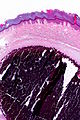
CC - very low mag.
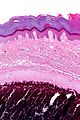
CC - low mag.
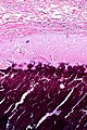
CC - intermed. mag.


www:
Sign out
SKIN AND SUBCUTANEOUS LESION, LEFT HIP, EXCISION:
- SUBCUTANEOUS CALCIFICATION SURROUNDED BY BENIGN FIBROUS TISSUE.
- DERMAL SCAR.
- NEGATIVE FOR MALIGNANCY.
SUBCUTANEOUS MASS, OVER BURSA OF ELBOW, EXCISION:
- CALCINOSIS CUTIS.
Micro
The sections show calcifications surrounded by macrophages and giant cells. No nuclear atypia is apparent. The overlying epidermis is unremarkable.
Without epidermis
The sections show dermal/subcutaneous calcifications surrounded by fibrosis, macrophages and giant cells. No nuclear atypia is apparent. Overlying epidermis is absent.
See also
References
- ↑ Dubey, S.; Sharma, R.; Maheshwari, V. (2010). "Scrotal calcinosis: idiopathic or dystrophic?". Dermatol Online J 16 (2): 5. PMID 20178701.
- ↑ URL: http://emedicine.medscape.com/article/1103137-overview. Accessed on: 21 September 2011.