Chorangioma
Jump to navigation
Jump to search
The printable version is no longer supported and may have rendering errors. Please update your browser bookmarks and please use the default browser print function instead.
| Chorangioma | |
|---|---|
| Diagnosis in short | |
 Chorangioma. H&E stain. | |
|
| |
| LM | abundant capillaries surrounded by stroma |
| LM DDx | chorangiosis, chorangiomatosis |
| Gross | white or red lesion |
| Site | placenta |
|
| |
| Associated Dx | +/-IUGR, +/-fetal hydrops - if large |
| Prevalence | uncommon |
Chorangioma is an uncommon pathology of the placenta that is similar to a hemangioma.
General
- Hamartoma-like growth in the placenta consisting of blood vessels.[1]
Epidemiology:
- Often benign/insignificant; large lesions (>4 cm[1] or >5 cm[2]) or multiple lesions are significant.
- May be association with:
- Fetal maternal haemorrhage.
- Fetal hydrops.
- IUGR.
- Incidence: ~1 in 100 placentas.[1]
Gross
- White lesions.
- Occasionally red lesions.
Microscopic
Features:[1]
- Mass of capillaries - key feature.
- +/-High cellularity.
- +/-Degenerative changes.
Notes:
- Must be differentiated from chorangiomatosis (associated with preeclampsia & IUGR) and chorangiosis (associated with maternal diabetes mellitus).[1]
Images
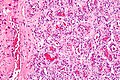
Chorangioma - high mag. (WC)
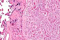
Chorangioma - intermed. mag. (WC)
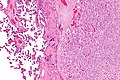
Chorangioma - low mag. (WC)


See also
References
- ↑ 1.0 1.1 1.2 1.3 1.4 Amer HZ, Heller DS (2010). "Chorangioma and related vascular lesions of the placenta--a review". Fetal Pediatr Pathol 29 (4): 199–206. doi:10.3109/15513815.2010.487009. PMID 20594143.
- ↑ Lež C, Fures R, Hrgovic Z, Belina S, Fajdic J, Münstedt K (2010). "Chorangioma placentae". Rare Tumors 2 (4): e67. doi:10.4081/rt.2010.e67. PMC 3019602. PMID 21234259. https://www.ncbi.nlm.nih.gov/pmc/articles/PMC3019602/.